
Abstract
The microconstituents of Ganoderma lucidum biomass (GLB) were evaluated, along with its antimicrobial and anticancer activities. Using gas chromatography-mass spectrometry analysis, 12-octadecadienoic acid (Z,Z)- and n-hexadecanoic acid with area% values of 21.0% and 11.0% were recognized in GLB. Uncooked biomass (UCB) and microwave-cooked (CE) biomass of G. lucidum caused a significant inhibition of human breast cancer (MCF-7) cell line proliferation in a dose-dependent manner. The inhibition of MCF-7 cell proliferation was 27.22 ± 1.64% using 16 µg/mL of CB while it was 52.29 ± 1.09% using 16 µg/mL of UCB. The cytotoxicity test recorded low IC50 (25.63 ± 0.52 µg/mL) of UCE compared to the IC50 value (49.99 ± 0.94 µg/mL) of CB. Highest antimicrobial activities were recorded via using UCE, compared to CE against Bacillus subtilis, Staphylococcus aureus, Escherichia coli, Klebsiella pneumoniae, Candida albicans, and Aspergillus niger. The 9,12-octadecadienoic acid (Z,Z)- and n-hexadecanoic acid were able to induce both anticancer and antibiotic-resistant properties. A key therapeutic target enzyme for evolving this resistant pathogenic activity on MCF-7 and K. pneumonia, was accessed thoroughly in silico study. The compounds described, therefore, might provide a great potential for the development of new therapeutics such as anticancer and antimicrobial agents.
Download PDF
Full Article
Characterization and Efficiency of Ganoderma lucidum Biomass as an Antimicrobial and Anticancer Agent
Mohammed Ibrahim Alghonaim,a Sulaiman A. Alsalamah,a Ahmed Alsolami,b,* and Tarek M. Abdel Ghany c,*
The microconstituents of Ganoderma lucidum biomass (GLB) were evaluated, along with its antimicrobial and anticancer activities. Using gas chromatography-mass spectrometry analysis, 12-octadecadienoic acid (Z,Z)- and n-hexadecanoic acid with area% values of 21.0% and 11.0% were recognized in GLB. Uncooked biomass (UCB) and microwave-cooked (CE) biomass of G. lucidum caused a significant inhibition of human breast cancer (MCF-7) cell line proliferation in a dose-dependent manner. The inhibition of MCF-7 cell proliferation was 27.22 ± 1.64% using 16 µg/mL of CB while it was 52.29 ± 1.09% using 16 µg/mL of UCB. The cytotoxicity test recorded low IC50 (25.63 ± 0.52 µg/mL) of UCE compared to the IC50 value (49.99 ± 0.94 µg/mL) of CB. Highest antimicrobial activities were recorded via using UCE, compared to CE against Bacillus subtilis, Staphylococcus aureus, Escherichia coli, Klebsiella pneumoniae, Candida albicans, and Aspergillus niger. The 9,12-octadecadienoic acid (Z,Z)- and n-hexadecanoic acid were able to induce both anticancer and antibiotic-resistant properties. A key therapeutic target enzyme for evolving this resistant pathogenic activity on MCF-7 and K. pneumonia, was accessed thoroughly in silico study. The compounds described, therefore, might provide a great potential for the development of new therapeutics such as anticancer and antimicrobial agents.
DOI: 10.15376/biores.18.4.8037-8061
Keywords: Lactate dehydrogenase; Breast cancer; Ganoderma lucidum; Antimicrobial activity
Contact information: a: Department of Biology, College of Science, Imam Mohammad Ibn Saud Islamic University, Riyadh 11623, Saudi Arabia; b: Department of Internal Medicine, College of Medicine, University of Ha’il, Hail 55476, Saudi Arabia; c: Botany and Microbiology Department, Faculty of Science, Al-Azhar University, Cairo 11725, Egypt;
*Corresponding author: tabdelghany.201@azhar.edu.eg; a.alsolami@uoh.edu.sa
GRAPHICAL ABSTRACT
INTRODUCTION
One of the primary causes of death worldwide, and a serious health issue, is cancer. Despite a noticeable rise in early diagnosis methods and advancements in treatment methods, cancer remains one of the most lethal illnesses in the world and presents a significant therapeutic challenge (Qanash et al. 2023a). Most governments are dedicated to reducing the harm that cancer poses to human health. The common cause of cancer-related fatalities among women globally is breast cancer (Su et al. 2018; Ghasaq et al. 2023). Today, the most frequent malignancy in women is breast cancer. Among advanced clinical studies, the major treatments for breast cancer are surgical removal, adjuvant chemotherapy, radiation, and hormone therapy. However, despite these restrictions, certain treatments’ efficacies remain unsatisfactory, particularly against triple-negative breast cancer, which does not respond to hormonal or trastuzumab-based therapy, as well as the rise in drug resistance and undesirable effects of synthetic drugs (Reddy 2011). Prevention of cancer proliferation via drug treatment, particularly natural compounds, has become an important health objective. It has become important to find anticancer medications with great effectiveness and minimal side effects. Thus, scientists have been working to identify bioactive components in natural resources. Numerous bioactive compounds, comprising anti-tumor drugs, were recently discovered in a variety of mushrooms (Zhong et al. 2019). Polysaccharides, lipids, proteins, ash, alkaloids, glycosides, volatile oils, phenolics, tocopherols, flavonoids, carotenoids, as well as organic acids are among the constituents of mushrooms that play a vital role in biological activities (Aparna et al. 2012; Sun et al. 2015; Joseph et al. 2018; Soliman et al. 2022). Asian epidemiological investigators suggested that mushrooms could indeed prevent the breast cancer (Shin et al. 2010).
Among mushrooms, G. lucidum is one of the most thoroughly studied mushrooms, especially in traditional Chinese medicine and other Asian folk medicine, as a functional food and chemo preventive agent (Oliveira et al. 2014). Several studies reported that G. lucidum could provide anticancer effects in addition to cancer cell-targeting techniques, such as cell cycle arrest and apoptosis induction (Dai et al. 2017; Park 2022), and migration inhibition with more immune improvement (Sun et al. 2015). In addition, a recent clinical study demonstrated the effectiveness of G. lucidum extracts. The cited study showed that the patient’s immune system was improved, and the side effects of chemotherapy and radiotherapy were lessened when using G. lucidum extracts along with standard therapies, such as chemotherapy, radiotherapy, and surgery (Zhong et al. 2019). The extract from G. lucidum has been shown to be cytotoxic to hepatoma, cervical cancer, and lung carcinoma (Ruan et al. 2014). Proliferation inhibition of MCF-7 was reported using G. lucidum extract (Vidhya and Devara 2011). Another study demonstrated that liver tumor and MCF-7 were inhibited by G. lucidum extract (Attoub et al. 2013). Lactate dehydrogenase (LDH) is a fascinating potential pharmacological target for the treatment of cancer among glycolysis enzymes. Because LDH is primarily found in cytosol, it can be released into the supernatant during cell damage or lysis. As a result, LDH is frequently used in vitro cell culture systems to assess cytotoxicity and cell number. The metabolic changes linked to breast cancer can be investigated using LDH, a useful diagnostic marker for the metabolic syndrome. In the pyruvate-reducing direction, breast cancer cells produce more LDH and express more genes than nearby normal cells (Ghasaq et al. 2023). Several indicators were associated with apoptosis of cancer cells (Riedl and Shi 2004). For instance, Feng et al. (2017) reported that caspase-3 activities increment is considered as a biomarker of apoptosis as well as a positive indicator for evaluate the efficacy of drugs in treatment of cancer. Moreover caspase-3 stimulates stress encouraged cancer cell proliferation, cellular migration, and angiogenesis of tumor (Zhou et al. 2018).
Moreover, several previous studies revealed that G. lucidum extract was effective against various bacteria, including Bacillus cereus, B. anthracis, B. subtilis, Staphylococcus aureus, Micrococcus luteus, Klebsiella oxytoca, K. pneumoniae, Escherichia coli, Proteus vulgaris, Serratia marcescens, Salmonella typhimurium, S. typhi, and S. tompson (Heleno et al. 2013; Abdullah et al. 2020). Recently in vitro, and in vivo studies by Soliman et al. (2022) demonstrated that G. lucidum extract has antibacterial activity particularly Methicillin-resistant Staphylococcus aureus. Various studies indicated that texture and chemical constituents might be changed as a result of cooking process, such as microwaving, pressure-cooking, griddling, frying, and steaming (Sun et al. 2014; Yuan et al. 2022) to evaluate the nutritional value but not for pharmacological value. The authors’ research with other published papers will contribute to over knowledge about the right way of management of cancer and multidrug resistance microorganisms by G. lucidum extract, as well as its utilization as an adjunct to prevent the chemotherapy effects.
G. lucidum is a vital pharmaceutical mushroom that has been applied in Oriental medicine for several years. As declared above and according to published papers, numerous secondary metabolites have been extracted and separated from G. lucidum. These compounds have various chemical formulas and possess several biological activities. The selection of suitable conditions for extraction of these metabolites is essential in order to determine their chemical contents, activities, and extracted quantities. Therefore, the current study aimed to assess the influence of cooked sample via microwave heating on the chemical constituents of G. lucidum extract and study its activity against human breast cancer cell line (MCF-7 cells) and pathogenic microorganisms to compare its activity with uncooked G. lucidum extract. In addition, the molecular docking interaction among the main detected compounds 9,12-octadecadienoic acid (z,z) and n-hexadecanoic acid on the extract with MCF-7 cells and Klebsiella pneumonia proteins was investigated.
EXPERIMENTAL
Materials
Preparation of Ganoderma lucidum extract sample
The G. lucidum product in fresh form was obtained from Hypermarket in Egypt.
The moisture content of the fresh fruiting bodies of the sample was calculated on a wet basis (72.24 ± 2.5 %) utilizing the oven at 80 °C for 36 h. The obtained sample was divided into two parts. The first part (50 g) was extracted with 200 mL of mixed solvents containing methanol, chloroform, and distilled water, (2:2:1 v/v/v); then the upper part of solvent containing the extract was removed, solvent was removed via rotary evaporator (40 °C), and the obtained extract was concentrated and weighted (Uncooked extract). The second part (50 g) was cooked without additional water by placing it on a glass plate and cooked using a home microwave oven for 1.5 min at 900 W pending tender. This was followed by extraction (Cooked extract), as mentioned in the first part. For further studies, the extracts were stored at -10 °C (Hamad et al. 2022).
GC-MS assay of G. lucidum constituents
The constituents of uncooked extract (UCE) and cooked extract (CE) were dissolved in dimethyl sulfoxide (DMSO) and were analyzed by gas chromatograph interface united with a mass spectrometer (GC-MS) model (Thermo-Scientific, Waltham, MA, USA; MS (ISQ Single Quadrupole Mass Spectrometer)). One mL/min of high quality, 99.99% helium was used as the carrier gas at a constant flow rate, while a split ratio of 1:100 was used to inject 1 μL of the extract into the gas chromatograph (GC), using the capillary column, which has dimensions of 30 m × 0.32 mm × 0.25 µm (TR5MS). The ion-source temperature was 280 °C, whereas the injector temperature was kept at 250 °C. The oven was set at 110 °C for 2 min and then increased gradually by 10 °C/min up to 200 °C/min, and one more increase by 5 °C/min to be extended to 280 °C/min, and finally held at 280 °C for 9 min. The mass spectra were recorded at 70 eV, and the chromatography procedure took 20 min. According to the average peak area of each discovered constituent to the total areas, the level percentage of each detected constituent was estimated. As previously mentioned, the GC-MS spectra were compared to the database made available by the National Institute of Standard and Technology (NIST) (https://www.nist.gov/) (Abdelghany et al. 2021; Al-Rajhi et al. 2022b).
Cytotoxicity and microscopic studies of cooked and uncooked G. lucidum extract against MCF-7 cell line propagation
Human breast cancer cell line (MCF-7 cells) obtained from the American Type Culture Collection (ATCC, Rockville, MD) were tested to see how the cells’ proliferation could be inhibited by CE and UCE samples of G. lucidum. The grown tested cells (5 × 10⁴) in Dulbecco’s modified Eagle’s medium (DMEM), which contained L-glutamine (1%) and gentamycin (50 g/L), were kept in culture under specific conditions (37 °C, a humid environment, and 5% of CO2) for 24 h. The growth medium containing different concentrations (2, 4, 8, 16, 32, 64, 128, 256, and 512 μg/mL) of the examined extracts was added to the wells; then the plates were incubated at certain conditions (37 °C, a humid environment, and 5% CO2). Through a microplate reader model SunRise (Tecan, Inc., Lyon, France), the viability of the cells was determined, using the MTT colorimetric method at 570 nm. The following formula was employed to determine the cytotoxicity percent (CY%):
![]() (1)
(1)
To determine the MCF-7 survival curve following treatment, the relationship between living cells and extract concentration was plotted. The quantity required to have toxic effects on 50% of whole cells was known as the 50% inhibitory concentration (IC50). Using GraphPad Prism software (San Diego, CA, USA), the IC50 was calculated from the graphic plots of the quantity response curve for each concentration. An inverted microscope (CKX41; Olympus, Tokyo, Japan) and digital camera were used to take pictures of the treated and untreated MCF-7 under the microscope (Abdelghany et al. 2023). Vinblastine sulfate (1 mM) was utilized as a drug control in cell culture wells. The DMSO as a negative control was applied also against cancer cells.
Assessment of lactate dehydrogenase (LDH) in MCF-7 cell line
A 24-well cell culture plate was cultured with 1.5 × 10⁵ cells per well and incubated for 12 h. Then, some cultured wells were exposed to different concentrations of CE and UCE of G. lucidum, while other wells were unexposed. All plates were incubated for two days. The collected supernatant was then added to a black 96-well culture plate (200 μL per well). Based on the manufacturer’s instructions, an LDH cytotoxicity test kit was used to assess LDH liberation. The absorbance in each well was measured utilizing an enzyme-linked immunoassay device at a wavelength of 450 nm (Zaqout et al. 2012).
Assay of caspase-3 in MCF-7 cell line
Based on the manufacturer’s instructions, caspase-3 was detected via colorimetric method using Caspase substrate Set Plus (BioVision, Milpitas, CA, USA) (Sharma et al. 2014). The MCF-7 cells at level 2 M 106/mL were incubated for one day followed by treatment with the different concentrations of UCE and CE of G. lucidum. Then, the treated and untreated MCF-7 cells were collected and centrifuged at 1000 rpm for 5 min to obtain cells pellet. Buffer was added to the cells pellet for cells lyses, and then kept in ice for 10 min. The lysate cells were centrifuged for 2 min at 5000 rpm. Then 50 µL of supernatant containing lysate cells were transmitted to a microplate. Each created reaction mixture needed 50 µL of 2x reaction buffer involved 10 Mm of D-1,4-dithiothreitol DTT. Five µL of caspase substrate (p-nitroaniline) was added to each well at the last step, and then the specimen was incubated at 37 °C for 2 h. The peptide cleavage by the caspase released the chromophore p-nitroaniline. At 405 nm wavelength, the absorbance of developed color was measured via reader of FLUOstar Omega microplate (BMG Labtech, Mannheim, Germany). Caspase enzymatic potential level was proportionately directly to the color reaction. The caspase-3 colorimetric assay kit was obtained from Abnova, Germany.
Antimicrobial activity cooked and uncooked G. lucidum extract
The tested microorganisms in the current study were bacteria and fungi, including Bacillus subtilis ATCC 6633, Staphylococcus aureus ATCC 6538, Escherichia coli ATCC 8739, Klebsiella pneumoniae (ATCC13883), Candida albicans ATCC 10231, and Aspergillus niger. The disc diffusion technique was used to evaluate the antimicrobial activity of CE and UCE of G. lucidum. After being propagated into BHI agar, the microorganisms were left to grow for 24 h at 37 °C (for bacteria) and for 3 days at 30 °C (for fungi). The cellular suspension (McFarland 0.5, 1.5 x 10⁸ UFC/mL) was then grown on Muller-Hinton agar plates and incubated at 37 °C for 24 h. Gentamicin (5 µL, 100 g/mL) was added to the discs’ surface as control. The disc was loaded with DMSO, then dried and used as a negative control in disc diffusion technique. Zones of halo inhibition (mm) were used to express the results of antimicrobial activity (Al-Rajhi et al. 2022a).
Molecular docking study
Three D structures of breast cancer cell line and Klebsiella pneumonia were obtained from the online database Protein Data Bank (http://www.rcsb.org/pdb) with an access code PDB: 2LWL and 5ZGN, respectively. Two D structures of 9,12-octadecadienoic acid (z,z) and n-hexadecanoic acid were drawn using ChemDraw Ultra 15.0 and saved as MDL files (.sdf) for MOE. Hardware was applied for calculation, molecule modeling, and docking molecule personal computer (Dell Core i7 processor 1.99 GHz, 16 GB memory with Windows 10, 64-bit operating system). The software was applied, using Molecular Operating Environment (MOE 2019). The Department of Chemistry, Faculty of Mathematics and Natural Science at Padjadjaran University was granted the license for MOE 2019.0102 (Chemical Computing Group www.chemcomp.com) to create protein and ligand structures. While applying for, creating, and simulating the process, docking was the program MOE dock. Program Alpha Site Finder was used for predicting the position and binding pocket volume of the site of the ligand’s interaction with macromolecules. Preparation of target proteins are detailed below:
1. The co-ligand and water molecules from the protein’s active site were eliminated.
2. The hydrogen atoms were added with their typical shape to the structure.
3. The dummy sites served as the binding pocket by generating the active binding sites created by the MOE site finder.
4. The obtained pocket was saved as MOE to predict the ligand-protein.
Preparation of ligands
Through using the MMFF94 force field, the native ligands were reduced to their lowest energy state. Following 3D protonation and the rectification procedure, the final form was produced. The ligands were inserted into the site, using the triangle matcher approach after the general docking scenario was run for 100 ns on the stiff receptor atoms. The GBVI/WSA dG procedures for rescoring were used, along with the London dG as a scoring function. The binding free energy (S kcal/mol) and hydrogen bonds between substances and protein-containing amino acids with lengths under 3.5 A were used to rank the top five poses. Additionally, the results pose-with-pose in the co-crystal ligand position and before and after amendment, respectively, was compared using the RMSD and RMSD-refine fields (Qanash et al. 2022; Yahya et al. 2022).
Statistical analysis
All measurements were performed in triplicate to calculate the ± standard deviation (SD) and variance via SPSS ver. 22.0 software (IBM, Armonk, NY, USA).
RESULTS AND DISCUSSION
Detected Constituents of G. lucidum Extract via GC-MS
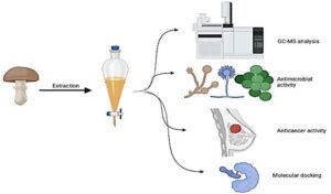
The CE and UCE of G. lucidum were subjected to GC-MS analysis and various biological activities (Fig. 1). Several compounds were detected with different retention time and area (%) (Tables 1 and 2, Figs. 2A and 2B) in CE and UCE of G. lucidum. The 9,12-octadecadienoic acid (Z,Z)- and n-hexadecanoic acid represented the greatest detected compounds with 20.99% and 10.97 %, respectively, while moderate area (%) was recognized for linoleic acid ethyl ester (7.01%) and propanoic acid, 2-(3-acetoxy-4,4,14-trimethylandrost-8-en-17-yl)- (4.67%) in UCE of G. lucidum. Moreover, cholestan-3-ol, 2-methylene-, (3á,5à)- and docosanoic acid, 1,2,3-propanetriyl ester were detected with the lowest area (0.92%) (Table 1). The GC-MS analysis showed differences in the numbers and area % of the recognized compounds of CE compared to UCE of G. lucidum. There were less numbers of the detected compounds in the CE than UCE of G. lucidum (Table 2), where several compounds, such as betulin, á-sitosterol, neoergosterone, glycidyl (Z)-9-heptadecenoate, and 2-bromotetradecanoic acid among others, were detected in UCE of G. lucidum. In contrast, some compounds were not affected by the coking process, such as octadecanoic acid and n-hexadecanoic acid. At the same time, some compounds were not detected in CE but appeared in UCE of G. lucidum; these probably were generated via transformation of original compound. The most detected constituents exhibited various biological activities similar to other studies. For example, n-hexadecanoic acid exhibited anti-inflammatory activity via inhibition of phospholipase A2, an anti-inflammatory enzyme (Aparna 2012). Moreover, n-hexadecanoic acid revealed anticancer potential against HCT-116 cells with excellent IC50 value (0.8 µg/mL) (Lokesh and Kannabiran 2017). Tian et al. (2018) reported that antioxidant activities were associated with the natural extracts, containing (Z,Z)-9,12-octadecadienoic acid (linoleic acid). Kusumah et al. (2020) documented the antibacterial activities of linoleic acid and α-linolenic acid against B. subtilis and S. aureus. As previously reported, several fatty acids could possess antifungal and antibacterial properties (Agoramoorthy et al. 2007); furthermore, fatty acids could regulate the immune responses by acting directly on T cells. The present results indicated the presence of n-hexadecanoic acid and 9,12-octadecadienoic acid (Z,Z)- in the extract of G. lucidum with high area %. According to Ohiri and Bassey (2016), n-hexadecanoic acid and 9,12-octadecadienoic acid (Z,Z)- represent the predominant constituents of G. lucidum that were detected via GC/MS analysis. Therefore these compounds were selected for molecular docking studies.

Fig. 1. Extraction method and various applicable tests to detect the constituents, antimicrobial activity, anticancer activity, and molecular docking of G. lucidum. This Image was designed via BioRender.com.

A

B
Fig. 2. GC-MS chromatogram analysis of UCE (A) and CE (B) samples of G. lucidum
Table 1. Detected Compounds of UCE of G. lucidum Assessed by GC-MS

Retention time (R.T.); molecular formula (M.F.), molecular weight (M.W.), the same is true for Table 2
Table 2. Detected Compounds of CE of G. lucidum Assessed by GC-MS

Inhibitory activity of cooked and uncooked G. lucidum extract against MCF-7 cancer cell line with morphological deviations
The MCF-7 cell line when exposed to different concentrations of CE and UCE of G. lucidum showed increment inhibition (%) of cell proliferation in a dose-dependent manner (Fig. 3). However, UCE exhibited more cytotoxicity against MCF-7 cell line than CE, where UCE at 2 µg/mL showed a significant inhibition (3.71 ± 0.16%) of cell proliferation, but at the same concentration of UCE, no cytotoxicity was reported. At 16 µg/mL, the inhibition of cells using CE and UCE was 27.22 ± 1.64% and 52.29 ± 1.09%, respectively. However, at high concentration of 512 µg/mL, the cell inhibition was approximately similar with slight differences for cells treated with UCE (87.56 ± 0.39%) and CE (88.66 ± 0.31%). Vinblastine sulfate, a positive drug of cancer treatment was used to evaluate the potential application of the extracts against MCF-7 cell line. IC50 was 25.63 ± 0.52 µg/mL for UCE and 49.99 ± 0.94 µg/mL for CE of G. lucidum extract, compared to the IC50 (6.87 ± 0.43 µg/mL) of vinblastine sulfate. Different extracts of mushrooms exhibited anticancer activities against MCF-7 cell line and other cancer cells with different activities. Kolniak-Ostek et al. (2022) has shown the potential cytotoxicity toward MCF-7 cell line of G. lucidum extract with IC50 209.6 µg/mL.

Fig. 3. Anticancer activities of CE and UCE samples against MCF-7 cell line
Cytotoxic effects of G. lucidum metabolites were reported against different cancer cell lines including breast, liver, lung, and, colon, due to the immunomodulatory properties of these metabolites (Cör Andrejč et al. 2022). However, other investigations are still required to further explain the immunomodulatory effect mechanisms in addition to the anticancer activities of G. lucidum. Recently, Kolniak-Ostek et al. (2022) tested the cytotoxicity of G. lucidum extract against tested cancer cell lines and normal cells. They demonstrated the extract exhibited cytotoxic effects on tumor cells and did not displayed toxicity against normal cells, which reveals a particular antitumor potential. From results of Sharmila et al. (2021), they demonstrated the successful application of G. Lucidum extract as anticancer agent because exhibited greater inhibition and mild activity towards breast cancer cell line and normal cells, respectively. A previous study reported the anticancer activity of Flammulina velutipes extract toward MCF-7 and MDA-MB-231 with different IC50 (17.7 to 38.36 µg/mL and 114.5 to184.2 µg/mL, respectively) values (Ukaegbua et al. 2018). The extracted polysaccharides of Agaricus bisporus displayed a strong block of MCF-7 cell growth but showed negligible cytotoxicity against colon, prostate, and gastric cancer (Jeong et al. 2012). Appropriately, colon and MCF-7 cell lines proliferation were affected by Pleurotus ostreatus extract with marked changes in the cell morphology, accompanied with the elongation in shape of MCF-7 cells (Jedinak and Sliva 2008; Mishra et al. 2022). In addition, the inhibitory mechanism of G. lucidum extract on MCF-7 cell lines included apoptosis induction, vitality reducing, and controlling key signaling molecules (Suarez-Arroyo et al. 2016). In the current findings, the differences among the activities of CE and UCE may be due to loss or transformation of active ingredients to other inactive ingredients because of cooking process. These observations were recorded in previous studies, but the antioxidant activity was investigated. Barros et al. (2007) claimed that the cooking of mushrooms using heat could damage the phenolics constructions, followed by declining their contents with a great antioxidant action into other phenolic constituents or altering phenolic constituents with little antioxidant action. Sun et al. (2014) also documented the low antioxidant activity that might be due to cooking process as not only reducing the phenolic compounds, but also altering the type and relative quantity of phenolics. The DMSO as a negative control did not exhibit toxicity against cancer cells.
MCF-7 cell line lactate dehydrogenase
According to several reports, the release amount of lactate dehydrogenase (LDH) represents one of the biomarkers to indicate the cancer cell destruction and apoptosis (Rose et al. 1993; Lai et al. 2008; Van Wilpe et al. 2020). In the current study, releasing LDH from MCF-7 cell line confirmed that UCE exerted more effect than CE (Fig. 4). Therefore, the quantity of the released LDH was high in cells exposed to UCE at different concentrations than its quantity when exposed to CE. The released amount of LDH was 16 and 9 Unit/mL at 256 µg/mL, and 13 and 7 Unit/mL at 512 µg/mL of UCE and CE, respectively.
Untreated cells released a higher quantity of LDH than treated; these cells indicated that of the tested extracts either UCE or CE affect the number of tumor cells. Several studies demonstrated that various tumors reveal high LDH expression (Buchakjian and Kornbluth 2010; Levine et al. 2010; Feng et al. 2018), which was attributed to bio-characteristics of malignant cells. These studies supported the authors’ explanation.

Fig. 4. MCF-7 cell line lactate dehydrogenase exposed to different concentrations of UCE and CE of G. lucidum
Activity of caspase-3 in exposed MCF-7 cells to UCE and CE of G. lucidum
Zhou et al. (2018) reported that caspase-3 is a main mediator of apoptosis stimulated during exposure of the cells to cytotoxic compounds, immunotherapy, or radiotherapy. It is often utilized as an indicator for the effectiveness of cancer therapy. In the current study, caspase-3 assay showed that the activity of caspase-3 increased in MCF-7 cells exposed to the different concentrations of UCE and CE of G. lucidum in a dose-dependent manner (Fig. 5). At all applied concentrations, the activity of caspase-3 was highest using UCE compared to the activity using the same concentration of CE, confirming the efficacy of UCE against MCF-7 cells. However, a brisk increment was observed in the activity of caspase-3 with increasing the concentration of the extracts, but exposure to 256 µg/mL of UCE and CE of G. lucidum displayed the major rise in the activity of caspase-3. Then, a slight increment in the activity was observed at 512 µg/mL. The results from this study were in line with other reports (Buchakjian and Kornbluth 2010), where in the other report the extract of Teucrium mascatense exhibited activity against MCF-7 cells. Caspase pathways are appropriate candidate objectives to find the drugs specific for cancer treatment as mentioned previously (Riedl et al. 2004). Generally, these findings support the findings that G. lucidum extract contains bioactive compounds that can cause death of MCF-7 cells via caspase-dependent apoptosis.

Fig. 5. Effect of UCE and CE of G. lucidum on the activity of Caspase-3 in MCF-7 cells
Antimicrobial activity of cooked and uncooked G. lucidum extract
Different species of bacteria and fungi were subjected to the CE and UCE of G. lucidum to evaluate the antimicrobial activity. It was clear that UCE provided antibacterial and antifungal activities more than CE against all tested microorganisms with different degree of inhibition zones (Fig. 6). Bacillus subtilis was the most sensitive bacteria with inhibition zone of 21 mm and 15 mm, while K. pneumonia was the most resistant bacteria with inhibition zones of 17 mm and 13 mm to UCE and CE, respectively. Promising anticandidal activity was recorded through using UCE and CE extracts against C. albicans with 20 mm and 17 mm inhibition zones, respectively. In contrast, both extracts exhibited less inhibitory action against A. niger with 8 mm and 6 mm inhibition zones, respectively. All obtained antimicrobial activities were compared utilizing standards of antibiotic and antifungal compounds (Fig. 7). As discussed in the anticancer activity of CE and UCE of G. lucidum, differences in the antimicrobial activity may be dependent on the components of the extract that were affected by the cooking procedures. Recently, Yuan et al. (2022) contributed to the number of detected volatile flavor compounds (54, 61, 53, 63, and 49 compounds) and the type of cooking methods (raw, steamed, boiled, microwaved, and fried) of Clitocybe squamulosa samples (Zhou et al. 2018). In the current study, the disc loaded with DMSO did not indicate any antimicrobial activity.

Fig. 6. Antimicrobial activities of CE and UCE G. lucidum extracts
Molecular docking study of 9,12-octadecadienoic acid (Z,Z) and n-hexadecanoic acid with MCF-7 cancer cell line 2LWL and K. pneumonia 5ZGN
The 9,12-octadecadienoic acid (Z,Z) and n-hexadecanoic acid were investigated to evaluate the binding affinities of MCF-7 cell line (2LWL) and K. pneumonia (5ZGN). This study observed that docking the compounds under study had higher binding scores against active sites of 5ZGN (-7.09431 kcal/mol and -6.65358 kcal/mol respectively) than that of 2LWL (-5.95503 kcal/mol and-5.68074 kcal/mol). 9,12-Octadecadienoic acid (Z,Z) was bound to the binding cavity of breast cancer (2LWL) with the residues (ASP-3 and PHE-2) via O-1, O-48, and C-32, respectively. Meanwhile, interacting with K. pneumonia (5ZGN) by (GLY-107 and PHE-144) via O-48 and C-19, respectively was documented. In contrast, n-hexadecanoic acid formed two hydrogen donor interactions against 2LWL with the residues of ASP-3 via O-1 and C-4. The interaction between n-hexadecanoic acid and the active site bound of 5ZN also revealed the presence of three acceptor hydrogen atoms between GLN-106, GLY-107, and TRP-108 amino acid residues and O-46 atom in the ligand. Several hydrogen bonds that existed between the identified proteins and the selected compounds were detected (Tables 3 and 4). In addition, the interaction of 9,12-octadecadienoic acid (Z,Z) and n-hexadecanoic acid with 2LWL and 5ZGN proteins was reported (Tables 5 and 6). The compounds docked to adopt their best-fitted postures (Figs. 7 and 8).
Some studies compared between biological activities of detected active constituents via molecular docking, for example Gupte et al. (2018) studied the molecular docking of four compounds including ganoderal A, ganoderol A, ganoderol B, and ganoderic acid Y as a natural constituents of G. lucidum with Lanosterol 14 α-demethylase enzyme as well as the inhibitory action of ganomycin1 and 2 on HIV 1 protease and Tyrosinase. The docking findings reflected the inhibitory potential of these constituents, demonstrating the probability that they can be utilized as potent drugs in the future. In another study, the docking interaction tool was performed to compare between interaction of ellagic acid and chlorogenic acid with the crystal structures of pathogenic yeasts including C. albicans (4YDE) and G. candidum (4ZZT). The obtained results indicated that chlorogenic acid was more active with a docking score of −7.84379 kcal/mol, than ellagic acid with a docking score of −6.18615 kcal/mol (Alsalamah et al. 2023). Recently, in silico studies on interaction of P. ostreatus active constituents with MCF-7 cancer cell line was documented (Mishra et al. 2022). Several scientific reports about docking scores were described in various reports studied, confirming the effectiveness of natural constituents from plants and fungi to repress the proliferation of different cancer cells as well as the inhibition of human pathogenic microbes (Al-Rajhi et al. 2022c,d,e; Al-Rajhi et al. 2023; Qanash et al. 2023b).


Fig. 7. Molecular docking processes of 9,12-octadecadienoic acid (Z,Z) and n-hexadecanoic acid with 2LWL protein


Fig. 8. Molecular docking processes of 9,12-octadecadienoic acid (Z, Z) and n-hexadecanoic acid with 5ZGN protein
Table 3. Docking Score and Energies of 9,12-Octadecadienoic Acid (z,z) and n-Hexadecanoic Acid with 2LWL Receptors

Table 4. Docking Score and Energies of 9,12-Octadecadienoic Acid (Z,Z) and n-Hexadecanoic Acid with 5ZGN Receptors

Table 5. 9,12-Octadecadienoic Acid (Z,Z) and n-Hexadecanoic Acid Interactions with 2LWL Protein

Table 6. 9,12-Octadecadienoic Acid (Z,Z) and n-Hexadecanoic Acid Interactions with 5ZGN Protein

CONCLUSIONS
- Various constituents with different biological activities were detected in the Ganoderma lucidum biomass (GLB).
- Increment in the inhibition (%) of MCF-7 cell line proliferation was recorded, using CE and UCE of GLB; however, UCE showed higher cytotoxicity than CE.
- The released quantity of LDH from treated MCF-7 cell line was more than the cells exposed to CE. Moreover, it might be concluded that G. lucidum extract, particularly UCE, might inhibit the proliferation and development of MCF-7 cells via stimulation of apoptosis by caspase-dependent pathways.
- The tested microorganisms were more affected by UCE compared to CE.
- MOE 2019.0102 is applicable to predict the best conformer of ligand structure and investigate the binding free energies of these inhibitors inside the target receptor. In this study, the docked 9,12-octadecadienoic acid (Z,Z) and n-hexadecanoic acid structural activities in silico with breast cancer cell line (2LWL) and K. pneumonia (5ZGN) drug target enzyme were reported.
FUNDING
This work was supported and funded by the Deanship of Scientific Research at Imam Mohammad Ibn Saud Islamic University (IMSIU) (grant number IMSIU-RP23038), Riyadh, Saudi Arabia.
ACKNOWLEDGMENTS
The authors wish to appreciate the Deanship of Scientific Research at Imam Mohammad Ibn Saud Islamic University (IMSIU) for support and funding the current study (grant number IMSIU-RP23038)
REFERENCES CITED
Abdelghany, T. M., Al-Rajhi, A. M. H., Yahya, R., Bakri, M. M., Al Abboud, M. A., Yahya, R., Qanash, H., Bazaid, A. S., and Salem, S. S. (2023). “Phytofabrication of zinc oxide nanoparticles with advanced characterization and its antioxidant, anticancer, and antimicrobial activity against pathogenic microorganisms,” Biomass Conversion and Biorefinery 13, 417-430. DOI: 10.1007/s13399-022-03412-1
Abdelghany, T. M., Yahya, R., Bakri, M. M., Ganash, M., Amin, B. H., and Qanash, H. (2021). “Effect of Thevetia peruviana seeds extract for microbial pathogens and cancer control,” Int. J. Pharmacol. 17(8), 643-655. DOI: 10.3923/ijp.2021.643.655
Abdullah, S., Jang, S. E., Kwak, M. K., and Chong, K. P. (2020). “Ganoderma boninense mycelia for phytochemicals and secondary metabolites with antibacterial activity,” J. Microbiol. 58, 1054-1064. DOI: 10.1007/s12275-020-0208-z
Agoramoorthy, M., Chandrasekaran, V., and Venkatesalu, M. J. H. (2007). “Antibacterial and antifungal activities of fatty acid methyl esters of the blind-your-eye mangrove from India,” Braz. J. Microbiol. 38(4), 739-742. DOI: 10.1590/S1517-83822007000400028
Al-Rajhi, A. M. H., Mashraqi, A., Al Abboud, M. A., Shater, A.-R. M., Al Jaouni, S. K., Selim, S., and Abdelghany, T. M. (2022a). “Screening of bioactive compounds from endophytic marine-derived fungi in Saudi Arabia: Antimicrobial and anticancer potential,” Life 12(8), article 1182. DOI: 10.3390/life12081182
Al-Rajhi, A. M. H., Yahya, R., Bakri, M. M., Reham, Y., and Abdelghany, T. M. (2022b). “In situ green synthesis of Cu-doped ZnO based polymers nanocomposite with studying antimicrobial, antioxidant and anti-inflammatory activities,” Appl. Biol. Chem. 65, article 35. DOI: 10.1186/s13765-022-00702-0
Al-Rajhi, A. M. H., Qanash, H., Almuhayawi, M. S., Al Jaouni, S. K., Bakri, M. M., Ganash, M., Salama, H. M., Selim, S., and Abdelghany, T. M. (2022c). “Molecular interaction studies and phytochemical characterization of Mentha pulegium L. constituents with multiple biological utilities as antioxidant, antimicrobial, anticancer and anti-hemolytic agents,” Molecules 27(15), article 4824. DOI: 10.3390/molecules27154824
Al-Rajhi, A. M. H., Salem, S. S., Alharbi, A. A., and Abdelghany, T. M. (2022d). “Ecofriendly synthesis of silver nanoparticles using Kei-apple (Dovyalis caffra) fruit and their efficacy against cancer cells and clinical pathogenic microorganisms,” Arab. J. Chem. 15(7), article ID 103927. DOI: 10.1016/j.arabjc.2022.103927
Al-Rajhi, A. M. H., Yahya, R., Abdelghany, T. M., Fareid, M. A., Mohamed, A. M., Amin, B. H., and Masrahi, A. S. (2022e). “Anticancer, anticoagulant, antioxidant and antimicrobial activities of Thevetia peruviana latex with molecular docking of antimicrobial and anticancer activities,” Molecules 27(10), article 3165. DOI: 10.3390/molecules27103165
Al-Rajhi, A. M. H., and Abdel Ghany, T. M. (2023). “Nanoemulsions of some edible oils and their antimicrobial, antioxidant, and anti-hemolytic activities,” BioResources 18(1), 1465-1481. DOI: 10.15376/biores.18.1.1465-1481
Alsalamah, S. A., Alghonaim, M. I., Jusstaniah, M., and Abdelghany, T. M. (2023). “Anti-yeasts, antioxidant and healing properties of henna pre-treated by moist heat and molecular docking of its major constituents, chlorogenic and ellagic acids, with Candida albicans and Geotrichum candidum proteins,” Life 13(9), article 1839. DOI: 10.3390/life13091839
Aparna, V., Dileep, K.V., Mandal, P. K., Karthe, P., Sadasivan, C., and Haridas, M. (2012). “Anti-inflammatory property of n-hexadecanoic acid: Structural evidence and kinetic assessment,” Chem. Biol. Drug Des. 80(3), 434-439. DOI: 10.1111/j.1747-0285.2012.01418.x
Attoub, S., Sperandio, O., Raza, H., Arafat, K., Al-Salam, S., Al Sultan, M. A., Al Safi, M., Takahashi, T., and Adem, A. (2013). “Thymoquinone as an anticancer agent: Evidence from inhibition of cancer cells viability and invasion in vitro and tumor growth in vivo,” Fund. Clin. Pharmacol. 27(5), 557-569. DOI: 10.1111/j.1472-8206.2012.01056.x
Barros, L., Baptista, P., Correia, D. M., Morais, J. S., and Perreira, I. C. R. (2007). “Effects of conservation treatment and cooking on the chemical composition and antioxidant activity of Portuguese wild edible mushrooms,” J. Agric. Food. Chem. 55(12), 4781-4788. DOI: 10.1021/jf070407o
Buchakjian, M. R., and Kornbluth S. (2010). “The engine driving the ship: Metabolic steering of cell proliferation and death,” Nat. Rev. Mol. Cell. Biol. 11(10), 715-727. DOI: 10.1038/nrm2972
Cör Andrejč, D., Knez Ž., and Knez Marevci, M. (2022). “Antioxidant, antibacterial, antitumor, antifungal, antiviral, anti-inflammatory, and nevro-protective activity of Ganoderma lucidum: An overview,” Front Pharmacol. 22(13), article 934982. DOI: 10.3389/fphar.2022.934982.
Dai, J., Miller, M. A., Everetts, N. J., Wang, X., Li, P., Li, Y., Xu, J. H., and Yao, G. (2017). “Elimination of quiescent slow-cycling cells via reducing quiescence depth by natural compounds purified from Ganoderma lucidum,” Oncotarget 8, 13770-13781. DOI: 10.18632/oncotarget.14634
Feng, X., Yu, Y., He, S., Cheng, J., Gong, Y., Zhang, Z., Yang, X., Xu, B., Liu, X., Li, C. Y., et al. (2017). “Dying glioma cells establish a proangiogenic microenvironment through a caspase 3 dependent mechanism,” Cancer Lett. 385, 12-20. DOI: 10.1016/j.canlet.2016.10.042
Feng, Y., Xiong, Y., Qiao, T., Li, X., Jia, L., and Han, Y. (2018). “Lactate dehydrogenase A: A key player in carcinogenesis and potential target in cancer therapy,” Cancer Med. 7(12), 6124-6136. DOI: 10.1002/cam4.1820
Ghasaq, J. K., Baydaa, A. A., Layla, O. F., Isam, N. S., and Ashgan, S. D. (2023). “A comparative study to determine LDH enzyme levels in serum samples of women with breast cancer and women with breast cancer and type 2 diabetes mellitus,” J. Med. Chem. Sci. 6(4) 883-890.
Gupte, A., Palande, A., Venkata, S., and Pol, R. (2018). “Docking studies of Ganoderma lucidum,” Int. J. Pharm. Sci. Res. 9(3), 1100-1105. DOI: 10.13040 /IJPSR.0975-8232.9(3).1100-05.
Hamad, D., El-Sayed, H., Ahmed, W., Sonbol, H., and Ramadan, M. A. H. (2022). “GC-MS analysis of potentially volatile compounds of Pleurotus ostreatus polar extract: In vitro antimicrobial, cytotoxic, immunomodulatory, and antioxidant activities,” Front. Microbiol. 13, article ID 834525 DOI: 10.3389/fmicb.2022.834525
Heleno, S. A., Ferreira, I. C. F. R., Esteves, A. P., Ćirić, A., Glamočlija, J., Martins, A., Soković, M., and Queiroz, M. J. R. P. (2013). “Antimicrobial and demelanizing activity of Ganoderma lucidum extract, p-hydroxybenzoic and cinnamic acids and their synthetic acetylated glucuronide methyl esters,” Food. Chem. Toxicol. 58, 95-100. DOI: 10.1016/j.fct.2013.04.025
Jedinak, A., and Sliva, D. (2008). “Pleurotus ostreatus inhibits proliferation of human breast and colon cancer cells through p53-dependent as well as p53-independent pathway,” Int. J. Oncol. 33(6), 1307-1313.
Jeong, S. C., Koyyalamudi, S. R., and Pang, G. (2012). “Dietary intake of Agaricus bisporus white button mushroom accelerates salivary immunoglobulin A secretion in healthy volunteers,” Nutrition 28(5), 527-531. DOI: 10.1016/j.nut.2011.08.005
Joseph, T. P., Chanda, W., Padhiar, A. A., Batool, S., LiQun, S., Zhong, M., and Huang, M. A. (2018). “Preclinical evaluation of the antitumor activities of edible and medicinal mushrooms: A molecular insight,” Integr. Cancer Ther. 17(2), 200-209. DOI: 10.1177/1534735417736861
Kolniak-Ostek, J., Oszmia´nski, J., Szyjka, A., Moreira, H., and Barg, E. (2022). “Anticancer and antioxidant activities in Ganoderma lucidum wild mushrooms in poland, as well as their phenolic and triterpenoid compounds,” Int. J. Mol. Sci. 23(16), article 9359. DOI: 10.3390/ijms23169359
Kusumah, D., Wakui, M., Murakami, M., Xie, X., Yukihito, K., and Maeda, I. (2020). “Linoleic acid, α-linolenic acid, and monolinolenins as antibacterial substances in the heat-processed soybean fermented with Rhizopus oligosporus,” Biosci. Biotech. Bioch. 84(6), 1285-1290. DOI: 10.1080/09168451.2020.1731299
Lai, J. C. K., Lai, M. B., Sirisha, J., Vikas, V. D., Alok, B., Christopher, K. D., and Solomon, W. L. (2008). “Exposure to titanium dioxide and other metallic oxide nanoparticles induces cytotoxicity on human neural cells and fibroblasts,” Int. J. Nanomed. 3(4) 533-545. DOI: 10.2147%2Fijn.s3234
Levine, A. J., and Puzio-Kuter, A. M. 2010). “The control of the metabolic switch in cancers by oncogenes and tumor suppressor genes,” Science 3, 330(6009), 1340-1344. DOI: 10.1126/science.1193494
Lokesh, R., and Kannabiran, K. (2017). “Cytotoxic potential of N-hexadecanoic acid extracted from Kigelia pinnata leaves,” Asian Journal of Cell Biology 12, 20-27. DOI: 10.3923/ajcb.2017.20.27
Mishra, V., Tomar, S., Yadav, P., Vishwakarma, S., and Singh, M. P. (2022). “Elemental analysis, phytochemical screening and evaluation of antioxidant, antibacterial and anticancer activity of Pleurotus ostreatus through in vitro and in silico approaches,” Metabolites 12(09), article 821. DOI: 10.3390/ metabo12090821
Ohiri, R. C., and Bassey, E. E. (2016). “Gas chromatography−mass spectrometry analysis of constituent oil from lingzhi or reishi medicinal mushroom, Ganoderma lucidum (agaricomycetes), from Nigeria,” International Journal of Medicinal Mushrooms 18(4), 365-369. DOI: 10.1615/IntJMedMushrooms.v18.i4.100
Oliveira, M., Reis, F. S., Sousa, D., Tavares, C., Lima, R. T., Ferreira, I. C., Tiago, D., and Vasconcelos, M. H. (2014). “A methanolic extract of Ganoderma lucidum fruiting body inhibits the growth of a gastric cancer cell line and affects cellular autophagy and cell cycle,” Food Funct. 5, 1389-1394. DOI: 10.1039/C4FO00258J
Park, H. J. (2022). “Current uses of mushrooms in cancer treatment and their anticancer mechanisms,” Int. J. Mol. Sci. 23(18), article 10502. DOI: 10.3390/ijms231810502
Qanash, H., Alotaibi, K., Aldarhami, A., Bazaid, A. S., Ganash, M., Saeedi, N. H., and Abdel Ghany, T. M. (2023a). “Effectiveness of oil-based nanoemulsions with molecular docking of its antimicrobial potential,” BioResources 18(1), 1554-1576. DOI: 10.15376/biores.18.1.1554-1576
Qanash, H., Bazaid, A. S., Aldarhami, A., Alharbi, B., Almashjary, M. N., Hazzazi, M. S., Felemban, H. R., and Abdelghany, T. M. (2023b). “Phytochemical characterization and efficacy of Artemisia judaica extract loaded chitosan nanoparticles as inhibitors of cancer proliferation and microbial growth,” Polymers 15(02), article 391. DOI: 10.3390/polym15020391
Qanash, H., Yahya, R., Bakri, M. M., Bazaid, A. S., Qanash, S., Shater, A. F., and Abdelghany, T. M. (2022). “Anticancer, antioxidant, antiviral and antimicrobial activities of Kei Apple (Dovyalis caffra) fruit,” Sci. Rep. 12(1), article 5914. DOI: 10.1038/s41598-022-09993-1
Reddy, K. B. (2011). “Triple-negative breast cancers: An updated review on treatment options,” Curr. Oncol. 18, e173-e179. DOI: 10.3747/co.v18i4.738
Riedl, S., and Shi, Y. (2004). “Molecular mechanisms of caspase regulation during apoptosis,” Nat. Rev. Mol. Cell. Biol. 5, 897-907. DOI: 10.1038/nrm1496
Rose, K., Goldberg, M. P., and Choi, D. W. (1993). “Cytotoxicity in murine neocortical cell culture,” Methods. Toxicol. 1A, 46–60.
Ruan, W., Lim, A. H., Huang, L. G., and Popovich, D. G. (2014). “Extraction optimisation and isolation of triterpenoids from Ganoderma lucidum and their effect on human carcinoma cell growth,” Nat. Prod. Res. 28(24), 2264-2272.
Sharma, M. K., Jalewa, J., and Hölscher, C. (2014). “Neuroprotective and anti-apoptotic effects of liraglutide on SH-SY5Y cells exposed to methylglyoxal stress,” J. Neurochem. 128(3), 459-471. DOI: 10.1111/jnc.12469
Sharmila, R., Akila, K., Manigandan, G., and Jayaramanathan, V. (2021). “Anti-cancer activity of Ganoderma lucidum mycelium alcoholic extract against breast cancer cells,” Uttar Pradesh Journal of Zoology 42(24), 65-77.
Shin, A., Kim, J., Lim, S. Y., Kim, G., Sung, M. K., Lee, E. S., and Ro, J. (2010). “Dietary mushroom intake and the risk of breast cancer based on hormone receptor status,” Nutr. Cancer 62(4), 476-483. DOI: 10.1080/01635580903441212
Soliman, A. M., Younis, A. M., Abdelgany, T. M., and Abdelbary, S. (2022). “Trends in assessment of Ganoderma lucidum methanol extract against MRSA infection in vitro and in vivo with nutrition support,” J. Adv. Pharm. Res. 6(1), 45-57. DOI: 10.21608/aprh.2022.111305.1147
Su, J., Li, D., Chen, Q., Li, M., Su, L., Luo, T., Liang, D., Lai, G., Shuai, O., Jiao, C., et al. (2018). “Anti-breast cancer enhancement of a polysaccharide from spore of Ganoderma lucidum with paclitaxel: Suppression on tumor metabolism with gut microbiota reshaping,” Front. Microbiol. 9, article 3099. DOI: 10.3389/fmicb.2018.03099
Suarez-Arroyo, I. J., Rios-Fuller, T. J., Feliz-Mosquea, Y. R., Lacourt-Ventura, M., Leal-Alviarez, D. J., Maldonado-Martinez, G., Cubano, L. A., Martinez-Montemayor, M. M. (2016). “Ganoderma lucidum combined with the EGFR tyrosine kinase inhibitor, erlotinib synergize to reduce inflammatory breast cancer progression,” J. Cancer. 7, 500-551. DOI: 10.7150/jca.13599
Sun, L., Bai, X., and Zhuang Y. (2014). “Effect of different cooking methods on total phenolic contents and antioxidant activities of four Boletus mushrooms,” J. Food Sci. Technol. 51(11), 3362-3368. DOI: 10.1007/s13197-012-0827-4
Sun, L. X., Li, W. D., Lin, Z. B., Duan, X. S., Xing, E. H., Jiang, M. M., Yang, N., Qi, H. H., Sun, Y., Li, M., et al. (2015). “Cytokine production suppression by culture supernatant of B16F10 cells and amelioration by Ganoderma lucidum polysaccharides in activated lymphocytes,” Cell. Tissue Res. 360, 379-389. DOI: 10.1007/s00441-014-2083-6
Tian, C., Gao, X., Yang, J., Gua, Y., Wang, H., and Liu, M. (2018). “Chemical compositions, extraction technology and antioxidant activity of petroleum ether extract from Abutilon theophrasti Medic. leaves,” Int. J. Food. Prop. 21, 1789-1799. DOI: 10.1080/10942912.2018.1494198
Ukaegbua, C. I., Shaha, S. R., Hazrulrizawatia, A. H., and Alara, O. R. (2018). “Acetone extract of Flammulina velutipes caps: A promising source of antioxidant and anticancer agents,” Beni-Suef University Journal of Basic and Applied Sciences 7(4), 675-682. DOI: 10.1016/j.bjbas.2018.07.012
Van Wilpe, S., Koornstra, R., Brok, M. D., De Groot, J. W., Blank, C., De Vries, J., Gerritsen, W., and Mehra, N. (2020). “Lactate dehydrogenase: A marker of diminished antitumor immunity,” OncoImmunology 9(1), article ID 1731942. DOI: 10.1080/2162402X.2020.1731942
Vidhya, N., and Devaraj, S. N. (2011). “Induction of apoptosis by eugenol in human breast cancer cells,” Indian J. Exp. Biol. 49(11), 871-878.
Yahya, R., Al-Rajhi, A. M. H., Alzaid, S. Z., Al Abboud, M. A., Almuhayawi, M. S., Al Jaouni, S. K., Selim, S., Ismail, K. S., and Abdelghany, T. M. (2022). “Molecular docking and efficacy of Aloe vera gel based on chitosan nanoparticles against Helicobacter pylori and its antioxidant and anti-Inflammatory activities,” Polymers 14(15), article ID 2994. DOI: 10.3390/polym14152994
Yuan, H., Xu, L., Chang, M., Meng, J., Feng, C., Geng, X., Cheng, Y., and Liu, Z. (2022). “Effects of different cooking methods on volatile flavor compounds, nutritional constituents, and antioxidant activities of Clitocybe squamulosa,” Front. Nutr. 9, article ID 1017014. DOI: 10.3389/fnut.2022.1017014
Zaqout, M. S., Sumizawa, T., Igisu, H., Wilson, D., Myojo T., and Ueno, S. (2012). “Binding of titanium dioxide nanoparticles to lactate dehydrogenase,” Environ. Health Prev. Med. 17, 341-345. DOI: 10.1007/s12199-011-0245-7
Zhong, L., Yan, P., Lam, W.C., Yao, L., and Bian, Z. (2019). “Coriolus versicolor and Ganoderma lucidum related natural products as an adjunct therapy for cancers: A systematic review and meta-analysis of randomized controlled trials,” Front. Pharmacol. 10, article 703. DOI: 10.3389/fphar.2019.00703
Zhou, M., Liu, X., Li, Z., Huang, Q., Li, F., and Li, C. Y. (2018). “Caspase‐3 regulates the migration, invasion and metastasis of colon cancer cells,” Int. J. Cancer 143(4), 921-930. DOI: 10.1002/ijc.31374
Article submitted: August 4, 2023; Peer review completed: October 4, 2023; Revised version received: October 6, 2023; Accepted: October 7, 2023; Published: October 11, 2023.
DOI: 10.15376/biores.18.4.8037-8061