
Abstract
Recently, algae have attracted the attention of investigators as a renewable source of compounds that can contribute to nanoparticle creation. The use algae biomass to facilitate preparation of copper oxide nanoparticles (CuONPs), as well as their application, were the aims of the present study. High performance liquid chromatography analysis of algal biomass indicated the presence of daidzein (2550 µg/mL), followed by ellagic acid (596 µg/mL). Algal biomass extract was successful as a bio-reducing agent for CuONPs fabrication at different temperatures up to 50 °C. Transmission electron microscopy characterized the created CuONPs with average size 5 to 17 nm. The colony radius of M. anisopliae, T. harzianum, C. lunata, F. oxysporium, A. flavus, and A. terreus was 1.84 ± 0.08, 1.97 ± 0.03, 1.00 ± 0.08, 2.04 ± 0.03, 2.32 ± 0.06, and 2.42 ± 0.05 cm, respectively at 200 mg of CuONPs. CuONPs exhibited inhibition zones of 26, 23, 25, and 22 mm when tested against B. subtilis, E.coli, K. pneumoniae, and S. aureus, respectively. Methyl orange and methyl green dyes were degraded by CuONPs with percentages ranging from 9.5 to 63.7% and from 22.3 to 75.7% at 15 to 90 min, respectively. Therefore, the created CuONPs can be regarded as excellent candidates for controlling fungal/bacterial development and dyes degradation.
Download PDF
Full Article
Algal Biomass Extract as Mediator for Copper Oxide Nanoparticle Synthesis: Applications in Control of Fungal, Bacterial Growth, and Photocatalytic Degradations of Dyes
Sulaiman A. Alsalamah,a Mohammed Ibrahim Alghonaim,a Abeer M. Mohammad,b and Tarek M. Abdel Ghany c,*
Recently, algae have attracted the attention of investigators as a renewable source of compounds that can contribute to nanoparticle creation. The use algae biomass to facilitate preparation of copper oxide nanoparticles (CuONPs), as well as their application, were the aims of the present study. High performance liquid chromatography analysis of algal biomass indicated the presence of daidzein (2550 µg/mL), followed by ellagic acid (596 µg/mL). Algal biomass extract was successful as a bio-reducing agent for CuONPs fabrication at different temperatures up to 50 °C. Transmission electron microscopy characterized the created CuONPs with average size 5 to 17 nm. The colony radius of M. anisopliae, T. harzianum, C. lunata, F. oxysporium, A. flavus, and A. terreus was 1.84 ± 0.08, 1.97 ± 0.03, 1.00 ± 0.08, 2.04 ± 0.03, 2.32 ± 0.06, and 2.42 ± 0.05 cm, respectively at 200 mg of CuONPs. CuONPs exhibited inhibition zones of 26, 23, 25, and 22 mm when tested against B. subtilis, E.coli, K. pneumoniae, and S. aureus, respectively. Methyl orange and methyl green dyes were degraded by CuONPs with percentages ranging from 9.5 to 63.7% and from 22.3 to 75.7% at 15 to 90 min, respectively. Therefore, the created CuONPs can be regarded as excellent candidates for controlling fungal/bacterial development and dyes degradation.
DOI: 10.15376/biores.18.4.7474-7489
Keywords: Algae; Nanoparticles; Fungi; Control; Photo-catalytic; Dyes; Degradation
Contact information: a: Department of Biology, College of Science, Imam Mohammad Ibn Saud Islamic University, Riyadh 11623, Saudi Arabia; b: Biology Department, Faculty of Science, Jazan University, Jizan, 12482, Jazan, Saudi Arabia; c: Botany and Microbiology Department, Faculty of Science, Al-Azhar University, Cairo 11725, Egypt; *Corresponding author: tabdelghany.201@azhar.edu.eg
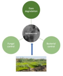
GRAPHICAL ABSTRACT
INTRODUCTION
The synthesis of eco-friendly nanoparticles (NPs) is an interesting area in nanobiotechnology. Biological processes to make NPs can be viewed as promising substitutes to chemical routes, as they have potential to avoid the generation of environmental impacting secondary pollutants. The green creation of NPs uses reductant agents from living organisms. Various biological creators of NPs, including algae, bacteria, fungi, and plants, have been documented in several studies (Abdelghany et al. 2018; Sharma and Kumar 2021; Al-Rajhi et al. 2022a; Abdelghany et al. 2023). These biological creators possess different molecules, such as polysaccharides, amino acids, alcoholic compounds, alkaloids, and vitamins responsible for stabilizing and reducing nanoparticles (Waris et al. 2021).
Algal biomass has been investigated as a prospective option for the environmentally friendly production of copper oxide nanoparticles (CuONPs). It is offered as a unique and simple process. The function of certain biomolecules and their contribution as capping and reluctant agents have not yet been fully investigated. The production of nanoparticles is made non-toxic and ecologically friendly using micro algae extracts as capping and reducing agents. When compared to other microorganisms, algae’s rapid growth and high biomass productivity have the added benefit of making the process more affordable. Algae may flourish in both clean water and effluent, which further enhances its environmental friendliness. Algae are being used extensively in the creation of nanoparticles. They are referred to as “bionanofactories” because they produce nanoparticles using both living and dead biomass of algae. Algae have a high capacity for absorbing metals, making the biological process that uses them economical and environmentally acceptable (Bilal et al. 2018). Large amounts of the reducing agent, which transforms metal salts into their corresponding metal nanoparticles without producing any harmful byproducts, are present in algae. Secondary metabolites, including polysaccharides, proteins, tannins, and steroids, are present in the aqueous extract of algae as bioactive compounds (Jin et al. 2016).
Because of their larger surface area per weight or volume and numerous characteristics, metal NPs have gained attention in recent years. Their thermal, biological, chemical, electrical, dielectric, physical, magnetic, mechanical, and optical properties make them appealing tools for research work (Khan et al. 2017). In a variety of areas, including medicine, screening, drug administration, antisensory, tissue biotechnology, cosmetics, gene engineering applications, and several others, nanoparticles play the most significant role (Sharma and Sharma 2017). Among the various metal oxide nanoparticles, copper oxide has received particular attention, as copper-based compounds possess effective biocidal properties and thus can be used in the formulations of pesticide and other health-related applications.
The antifungal activity of compounds containing copper has been reported and applied for a long time. It is still being applied today, even with its associated environmental problems. Many researchers recommend substitution of copper with CuONPs. Several fungi, including Fusarium culmorum, F. oxysporum, F. graminearum, F. solani, Aspergillus niger, A. flavus, Penicillium chrysogenum, and Alternaria alternata are inhibited by CuONPs (Shende et al. 2015; Abdelghany et al. 2020). Fungicidal activity of CuONPs was recorded on the genetic levels of Penicillium digitatum and F. solani (Khamis et al. 2017). Ultrastructure changes of F. incarnatum were recorded, such as injury of hypha, conidiospores, cell membranes, and walls, as result of treatment by CuONPs (Al-Rajhi et al. 2022b).
Several environmental problems arise and increase each year as a result of the discharge of dyes into water bodies (Mehra et al. 2021). Therefore, the search for new or development of approaches for dye degradation is a challenge for investigations (Abdelghany et al. 2019; Ihsanullah et al. 2020; Nemiwal et al. 2021; Qanash et al. 2023). Numerous semiconductors or metal oxides are efficient photocatalysts under different sources of light, because of their small band gaps. For instance, a small band gap ranging from 2.1 to 2.71 eV is associated with CuO (Karthikeyan et al. 2020). CuO NPs display an appropriate response towards mechanical, optical, and photolytic applications (Pourmoslemi et al. 2020). Algae are recognized for accumulating heavy metals and also have a remarkable capacity to transform them into more pliable forms. Because of these enticing characteristics, algae have been anticipated as model organisms for producing different forms of nanomaterials, particularly metallic NPs (Fawcett et al. 2017). Moreover, the environmental habitat of the collected biomass in the present study is rich with numerous minerals as well as the abundance of theses algae without any benefits. For these reasons, the algal biomass was used to create CuONPs with some applications including antifungal activity and dyes degradation.
EXPERIMENTAL
Source of Algal Biomass
Abundant green algae were developed in an agricultural drainage channel located at Monufia Governorate, Egypt (30° 62′8014″ N, 116° 31′ 070334″ E). This channel contains polluted water from different sources, including municipal, agricultural runoff, and industrial wastewater (Abdel Ghany et al. 2021). Fresh biomass (250 g) was collected from this channel and rinsed several times with distilled water. Then, the rinsed biomass was shade-dried, and 10 g of algal biomass were ground and immersed in 200 mL of distilled water, followed by simultaneously autoclaving and filtrating using filter paper (Whatman No. 1). Through centrifugations, the resulting supernatant from the filtered extract was utilized as the reducing agent for creating CuONPs.
Phenolic and flavonoid constituents of algal biomass
The washed and shade-dried algal biomass (50 g) was ground and extracted with 250 mL of methanol (20% w/v). The extract was subjected to high performance liquid chromatography (HPLC; Agilent 1260 Infinity HPLC Series, Agilent Technologies, Santa Clara, CA, USA) to detect the flavonoid and phenolic contents. The HPLC was fortified with a Quaternary pump and a Zorbax Eclipse amended with a column of C18 (100 mm × 4.6 mm i.d.). Twenty µL of the extract were injected in HPLC. Three gradient elutions were applied for the phenolic constituents separation at 30 °C including HPLC grade water 0.2% H3PO4 (v/v), methyl alcohol (B), and acetonitrile (C). The detector wavelength was applied at 284 nm. For flavonoid constituents’ separation, the Knauer HPLC was fortified by a binary pump, the applied gradient elutions consisted of methanol and 0.5% of H3PO4 in water (50:50 %) with 0.7 mL/min of flow rate. Twenty µL of algal extract were injected. The detector wavelength was applied at 284 nm for flavonoids detection. The identification of constituents was depended on the existence of standard constituents.
Biogenic synthesis of copper oxide nanoparticles.
With vigorous stirring for 1 day at 100 °C, 10 mL of algal extract (5% g/v) was added dropwise to 100 mL of 1 mM aqueous copper acetate in an Erlenmeyer flask (250 mL capacity) for synthesis of CuONPs as a positive reaction mixture. Moreover, copper acetate aqueous solution without algal extract was used as negative control and kept at the same conditions of the positive reaction mixture. If the color in positive reaction mixture was changed after 5 h from bright blue to dark brown, it was a sign that CuONPs were forming, but in the negative control, the color was unaltered. Regular color changes and UV-visible spectrum measurements were made to track the process’s development. The formed CuONPs were collected via centrifugation process for 15 min. The resulting CuONPs were then redispersed and cleaned by deionized H2O to remove any debris and uncoordinated biomolecules. This separation and washing procedure was repeated three times to ensure CuONPs separation. The obtained pure CuONPs were oven-dried to complete the characterization process (Shehabeldine et al. 2023).
Characterization of created CuONPs
The creation of CuONPs by algal biomass was documented via a UV-visible spectrophotometer (Nicolet evolution 100, Cambridge, MA, USA) in the wavelength range (200 to 700 nm). Additionally, the reaction mixture at different temperatures (30, 40, and 50 ℃) was evaluated using an UV-visible spectrophotometer for synthesis of CuONPs. Shape and size of created CuONPs were investigated using a transmission electron microscope (TEM; JEOL JEM-2100, Tokyo, Japan). The created CuONPs were suspended in aqueous solution; and then a suspension drop was transported onto the TEM grids, and then dried before examination. An X-ray diffractometer X’Pert Pro (Philips, Eindhoven, Netherlands) was applied to evaluate the crystallinity of CuONPs created by algal biomass. The temperature range of 2θ was 4 to 70 °C. The radiation of Ni-filtered Cu Ka was utilized as a source of X-ray, with 40 kV as voltage and 30 mA as current.
Antifungal and antibacterial activity
Various phytopathogenic (Curvularia lunata and Fusarium oxysporium), mycotoxigenic (Aspergillus flavus and Aspergillus terreus), and bio-applicable (Metarhizium anisopliae and Trichoderma harzianum) fungi were used for testing. Petri dishes contained a solid culture medium without tested compounds (CuONPs, copper acetate, algal extract, and copper oxychloride (chemical fungicide)), and a medium fortified with different concentrations of each tested compound (50, 100, and 200 mg/L). Fungal mycelia (6 mm of fungal disc) were transferred to the center of the agar plate’s surface, and then incubated at 30 °C for 7 days. Growth development of the tested fungi was estimated via measuring the colony radius compared to the control cultures (Abdelghany et al. 2015).
Nutrient agar plats were inoculated with tested bacteria including Staphylococcus aureus ATCC6538, Bacillus subtilis ATCC6633, Escherichia coli ATCC8739, and Klebsiella pneumoniae ATCC 8047 via a streaking method. Then, discs (6 mm) loaded with 100 µL of CuONPs, copper acetate, algal extract, and Gentamycin as standard antibiotic (20 µg/mL), were placed on the surface of inoculated agar with the tested bacteria. The plates were kept in refrigerator for 30 min to allow the diffusion of tested materials before bacterial growth, then incubated at 37 for 24 h, and then the appeared clear zones were measured (Humphries et al. 2018).
Photocatalytic test of CuONPs
According to Abdelghany and Al Abboud (2014) with some modification, the ability of CuONPs to degrade the methyl orange (MO) and methyl green (MG) dyes was estimated in the presence of visible light (500 km h−1m−2 as a mean solar flux). Each dye at concentration 10 mgL−1 was mixed with 10 mg of CuONPs. Then, for 30 min, the reaction mixture was agitated in the dark to authenticate the equilibrium of adsorption–desorption. A well-established spectrum of UV-vis absorption was seen in all experiments. Bands at 632 nm and 462 nm were scanned for MG and MO. The mixture reaction was then stirred under irradiation of sunlight, followed by withdrawing two mL of the reaction mixture each 15 min, up to 90 min, to detect the peak of absorption via UV-Vis at time “t”. To calculate the dye degradation, the following equation was used:

Statistical Assessment
One-way analysis of variance (ANOVA) in CoStat software (version 14, IBM Corp., Armonk, NY, USA) was applied to statistical studies, with significant differences detected via Tukey’s test post hoc besides standard deviation (SD) approaches. The differences in results with the similar letters are not significant.
RESULTS AND DISCUSSION
The water surface was covered with green algal biomass for a long time (Fig. 1A). In each period these algal biomass were removed (Fig. 1B) or disappeared as suitable conditions in the water were unavailable. The appearance of these algae is considered a bio-indicator of water content changes. Some kinds of algae develop in complex habitats, and they can live in extreme conditions such as temperature, salinity, ultraviolet radiation, and nutrients; therefore, to continue, they must adapt to stress conditions via producing various secondary metabolites (Sirbu et al. 2020). The HPLC analysis of algal biomass indicated that daidzein represented the most identified compounds with a concentration of 2550 µg/mL, followed by ellagic acid (596 µg/mL). Moderate concentrations were associated with caffeic acid (262 µg/mL), gallic acid (233 µg/mL), and chlorogenic acid (205 µg/mL). Some compounds were detected in very low concentrations, such as ferulic acid (7.93 µg/mL), cinnamic acid (18.6 µg/mL), methyl gallate (18.9 µg/mL), and apigenin 22.6 µg/mL). According to the availability of standard phenolics and flavonoids, five compounds not detected in algal biomass included catechin, syringic acid, pyro catechol, rutin, coumaric acid, vanillin, and hesperetin. In contrast, three compounds at different retention times were identified; however, these compounds occurred with high area (%) (Table 1 and Fig. 2). As mentioned, algae are a unique natural biomass as a source of a number of compounds with several valuable properties. Brodowska (2017) identified many constituents, such as daidzein, kaempferol, naringenin, and apigenin, in green algae. Quercetin, kaempferol, and naringenin were also detected in many algal extracts (Gentscheva et al. 2022).

Fig. 1. Collection site of algal biomass in agricultural drainage channel (A), agricultural drainage channel after cleaning from algae and wastes (B)

Fig. 2. Chromatograms of detected phenolic and flavonoid constituents via HPLC
Table 1. Phenolic and Flavonoid Constituents of Algal Biomass


As the algal extract contains numerous compounds of phenolic and flavonoids, as well as secondary metabolites, its ability to reduce copper compounds to nanoparticles increased. The main reducing agent in algae extract for CuONPs creation may be the compounds that possess several functional groups such as chlorogenic acid and quercetin. Therefore, the algal biomass was subjected to CuONPs synthesis. UV-visible spectroscopy showed the maximum absorption peak at 250 nm. Their surface plasmon resonance at different temperatures indicated that algal biomass was able to create CuONPs at elevated conditions of temperature (Fig. 3). The UV spectrum was compared to algal biomass without the addition of copper salt. The authors’ observation agrees with the other studies on the creation of CuONPs by algae, which shows that the CuONPs peak was at 247 nm (Mohamed et al. 2021a). The size and morphology of created CuONPs were characterized by TEM (Fig. 4). CuONPs appeared in spherical form (average size 5 to 17 nm). The dispersed NPs were surrounded with capping by algal active metabolites. Mohamed et al. (2021a) noted that the size range of created CuONPs was 21.8 nm. In this context, brown algae Macrocystis pyrifera was mediated for CuONPs creation within a size of 2 to 50 nm (Araya-Castro et al. 2021).

Fig. 3. Absorbance peaks of the green synthesized CuONPs using a UV – Vis spectrophotometer

Fig. 4. TEM micrograph of created CuONPs by algal biomass
The crystalline structure of CuONPs was validated using XRD analysis, as shown in Fig. 5. The primary strong angles in the diffractogram of biosynthesized CuONPs were visible in the XRD patterns, showing that CuONPs were crystallographic in nature (Mohamed et al. 2021b; Hammad et al. 2022). Figure 1 shows XRD diffraction peaks of CuONPs, and displays the diffraction characteristics regarding 2θ at 33.8°, 35.5°, 37.9°, 48.1°, 51.6°, 61.01°, and 66.2°, which represented the Bragg’s reflections at 110, 111, -111, -202, 020, -113, and 022, respectively. The mineral crystal was CuO of the tenorite crystal form. Thus, all of the observed peaks were similar to those reported by the joint committee on powder diffraction standards (JCPDS) of CuO-NPs with a standard card JCPDS File No: 01-1117, as recorded by Badawy et al. (2021), and Shehabeldine et al. (2023). Therefore, the results clearly support the CuONPs synthesis. The CuONPs diffractogram does not reveal the presence of any other impurities, ensuring that the CuONPs obtained were pure and in agreement with other investigations (Shende et al. 2015; Nabila and Kannabiran 2018; Mohamed et al. 2021).

Fig. 5. XRD pattern of the biosynthesized CuONPs
Copper compounds have been previously applied as fertilizer and fungicide, and several studies recommend that CuONPs up to certain concentrations are not toxic and efficiently repress disease development in plants (Faraz et al. 2022). The current investigation reflected the impact of CuONPs on phytopathogenic (C. lunata and F. oxysporium), mycotoxigenic (A. flavus and A. terreus), and bio-applicable (M. anisopliae and T. harzianum) fungi compared to copper acetate, algal extract, and chemical fungicide (Table 2). The growth of all tested fungi decreased with increasing concentration of CuONPs and fungicide, where at 50 mg the colony diameter was 4.02 ± 0.08, 5.92 ± 0.11, 4.50 ± 0.08, 7.10 ± 0.04, 4.78 ± 0.04, and 3.87 ± 0.03 cm; while at 200 mg the colony diameter was 1.84 ± 0.08, 1.97 ± 0.03, 1.00 ± 0.08, 2.04 ± 0.03, 2.32 ± 0.06, and 2.42 ± 0.05 cm of M. anisopliae, T. harzianum, C. lunata, F. oxysporium, A. flavus, and A. terreus, respectively. The chemical fungicide showed more inhibitory action than CuONPs. In contrast, algal extract exhibited stimulatory action for growth of all tested fungi at 50 and 100 mg, but at high concentration, 200 mg showed negligible fungal inhibition compared with the control.
Table 2. Effect of Different Concentrations of Copper Acetate, Algal Biomass, CuONPs and Chemical Fungicide on Fungal Growth


Growth of three fungi, including M. anisopliae, T. harzianum, and A. flavus, were encouraged with low concentration of 50 mg of copper acetate, where the colony growth was 6.81 ± 0.07, 7.84 ± 0.07, and 6.58 ± 0.04 cm compared to colony growth 6.78 ± 0.18, 7.80 ± 0.03, and 6.54 ± 0.03 cm for the control, respectively. The colony radius of C. lunata, F. oxysporium, and A.terreus decreased with increasing copper acetate. Finally, the inhibitory action against tested fungi was attributed to the chemical fungicide followed by CuONPs, followed by copper acetate, and algal extract. Banik and Pérez-de-Luque (2017) investigated the influence of CuONPs against phytopathogenic and beneficial microorganisms; they observed that CuONPs did not significantly inhibit Trichoderma harzianum and Rhizobium spp. compared to chemical fungicide (copper oxychloride). Additionally, they showed that the growth of Fusarium oxysporum, T. harzianum, Botrytis fabae, Alternaria alternate, and Pseudomonas syringae was promoted at low concentration of CuONPs. CuONPs interfere with germination of fungal spores via affecting the metabolic pathways of fungi (Gaba et al. 2022). Arya et al. (2018) documented the antibacterial and antifungal potential of created CuONPs by green alga Botryococcus braunii against Pseudomonas aeruginosa, Klebsiella pneumoniae, Escherichia coli, Staphylococcus aureus, Escherichia coli, and Fusarium oxysporum. Recently, Atri et al. (2023) showed that CuONPs reflected efficient antifungal activity.
Moreover, the antibacterial activity of CuONPs was recorded compared to to copper acetate, algal extract, and standard antibiotic (Fig. 6). CuONPs exhibited promising inhibitory action against all tested bacteria with inhibition zones of 26, 23, 25, and 22 mm compared to antibiotic with inhibition zones of 21, 22, 21, and 18 mm against B. subtilis, E. coli, K. pneumoniae, and S. aureus, respectively. Copper acetate also showed antibacterial activity against all tested bacteria but less CuONPs and antibiotic. Negligible inhibition zones were observed using algal biomass extract against only B. subtilis, and E. coli. According to Nabila and Kannabiran (2018), the green created CuONPs by actinomycetes revealed bacteriostatic potential against various bacterial pathogens for human and fish including Bacillus cereus, Edwardsiella tarda, S. aureus, Proteus mirabilis, Vibrio anguillarum, Aeromonas hydrophila, and A. caviae. Copper metal (Cu2+) exhibits activity against bacteria and is accepted by US-EPA (US-Environmental Protection Agency) as a harmless agent for fighting microorganisms. The CuONPs have more antibacterial activity compared to the non-nanoform of copper compound (Vasantharaj et al. 2023).

Fig. 6. Antibacterial activity of copper acetate, algal biomass, CuONPs, and antibiotic
Degradation quantity of methyl green and methyl orange dyes increased with the increasing time as a result of exposure to CuONPs as a photocatalyst (Fig. 7). At 90 min, the degradation quantity was 75.67 ± 0.58 and 64.67 ± 3.21% for methyl green and methyl orange dyes, respectively. There was negligible increase in degradation quantity of dyes after 75 min, where degradation quantity was 75.33 ± 1.53 and 63.67 ± 1.53% for methyl green at 75 min, while it was 75.67 ± 0.58 and 64.67 ± 3.21% at 90 min. If the exposure time was augmented, more efficiency of dyes degradation could be obtained. Degradation quantity of methyl orange was less than methyl green at all different times. Degradation of dyes by CuONPs was recorded in other studies but with different levels depending on many factors, such as dyes concentration as mentioned by Aroob et al. (2023), where 20 ppm of methyl green and methyl orange dyes showed less degradable compared to 10 ppm. Sharma and Sharma (2017) showed that methyl orange was degraded up to 96% via green synthesized CuONPs in the existence of UV light at 24 min, while Ikram et al. (2022) showed degradation efficiency was 45.2% and 32.0% via green synthesized CuONPs in the presence of UV light and sunlight, respectively, at 60 min. Atri et al. (2023) revealed that the green synthesized CuONPs can play a vital role in the degradation of dyes in industrial and domestic waste.

Fig. 7. Degradation quantity of methyl green and methyl orange by CuONPs at different times
CONCLUSIONS
- The available algae in the agricultural drainage channel were exploited in the green synthesis of copper oxide nanoparticles (CuONPs), and these NPs were applied to combat phytopathogenic and mycotoxigenic fungi, and their activity against fungi exploited in biotechnology was also evaluated compared to the effect of a chemical fungicide on all tested fungi.
- The created CuONPs, which were identified as tenorite based on X-ray diffraction (XRD), effectively revealed photocatalytic activity to remove methyl orange and methyl green degradation.
- The obtained promising findings offered attractive resources for investigators to produce economical and eco-friendly fungicide and photocatalyst to control fungal development and efficiently reduce dyes water contamination.
Funding
This work was supported and funded by the Deanship of Scientific Research at Imam Mohammad Ibn Saud Islamic University (IMSIU) (grant number IMSIU-RP23038), Riyadh, Saudi Arabia.
Acknowledgments
The authors wish to appreciate the Deanship of Scientific Research at Imam Mohammad Ibn Saud Islamic University (IMSIU) for support and funding the current study (grant number IMSIU-RP23038)
Conflicts of Interest
The authors declare no conflict of interest.
REFERENCES CITED
Abdel Ghany, T. M., Mahmoud, M. S., Alawlaqi, M. M., Reyad, A. M., Al-Rajhi, A. M. H., and Abdkareem, E. M. (2021). “Physicochemical characterization of agricultural run-off and groundwater inoculated by Trichoderma asperellum and its effect on anti-oxidative enzymes production by irrigated Trifolium alexandrinum L.,” BioResources 16(2), 3272-3284. DOI: 10.15376/biores.16.2.3272-3284
Abdelghany, T. M., and Al Abboud, M. A. (2014). “Capacity of growing, live and dead fungal biomass for safranin dye decolourization and their impact on fungal metabolites,” Aust. J. Basic Appl. Sci. 8(10), 489-499.
Abdelghany, T. M., Shater, A. R. M., Negm, M. E., Al Abboud, M. A., and Elhussieny, N. I. (2015). “Efficacy of botanical fungicides against Curvularia lunata at molecular levels,” J. Plant Pathol. Microb. 6, article 289. DOI: 10.4172/2157-7471.1000289
Abdelghany, T. M. A., Bakri, M. M., Al-Rajhi, A. M. H., Abboud, M. A. A., Alawlaqi, M. M., and Shater, A. R. M. (2020). “Impact of copper and its nanoparticles on growth, ultrastructure, and laccase production of Aspergillus niger using corn cobs wastes,” BioResources 15(2), 3289-3306. DOI: 10.15376/biores.15.2.3289-3306
Abdelghany, T. M., Al-Rajhi, A. M., Al Abboud, M. A., Alawlaqi, M. M., Ganash Magdah, A., Helmy, E. A., and Mabrouk, A. S. (2018). “Recent advances in green synthesis of silver nanoparticles and their applications: About future directions. A review,” BioNanoScience 8, 5-16. DOI: 10.1007/s12668-017-0413-3
Abdelghany, T., Abboud, M., Alawlaqi, M., and Shater, A. R. (2019). “Dead biomass of thermophilic Aspergillus fumigatus for Congo red biosorption,” The Egyptian Journal of Experimental Biology (Botany) 15(1), 1-6. DOI: 10.5455/egyjebb.20181206084342
Abdelghany, T. M., Al-Rajhi, A. M. H., Yahya, R., Bakri, M. M., Al Abboud, M. A., Yahya, R., Qanash, H., Abdulrahman, S. B., and Salem, S. S. (2023). “Phytofabrication of zinc oxide nanoparticles with advanced characterization and its antioxidant, anticancer, and antimicrobial activity against pathogenic microorganisms,” Biomass Conv. Bioref. 13, 417-430. DOI: 10.1007/s13399-022-03412-1
Al-Rajhi, A. M. H., Yahya, R., Bakri, M. M., Yahya R., and Abdelghany, T. M. (2022a). “In situ green synthesis of Cu-doped ZnO based polymers nanocomposite with studying antimicrobial, antioxidant and anti-inflammatory activities,” Appl. Biol. Chem. 65, article 35. DOI: 10.1186/s13765-022-00702-0
Al-Rajhi, A. M., Yahya, R., Alawlaqi, M. M., Fareid, M. A., Amin, B. H., and Abdelghany, T. M. (2022b). “Copper oxide nanoparticles as fungistat to inhibit mycotoxins and hydrolytic enzyme production by Fusarium incarnatum isolated from garlic biomass,” BioResources 17(2), 3042-3056. DOI: 10.15376/biores.17.2.3042-3056
Araya-Castro, K., Chao, T.-C., Durán-Vinet, B., Cisternas, C., Ciudad, G., and Rubilar, O. (2021). “Green synthesis of copper oxide nanoparticles using protein fractions from an aqueous extract of brown algae Macrocystis pyrifera,” Processes. 9(1), article 78. DOI: 10.3390/pr9010078
Aroob, S., Carabineiro, S. A. C., Taj, M. B., Bibi, I., Raheel, A., Javed, T., Yahya, R., Alelwani, W., Verpoort, F., Kamwilaisak, K., et al. (2023). “Green synthesis and photocatalytic dye degradation activity of CuO nanoparticles,” Catalysts 13(3), article 502. DOI: 10.3390/catal13030502
Arya, A., Gupta, K., Chundawat, T. S., and Vaya, D. (2018). “Biogenic synthesis of copper and silver nanoparticles using green alga Botryococcus braunii and its antimicrobial activity,” Bioinorg Chem Appl. 2018, article ID 7879403. DOI: 10.1155/2018/7879403
Atri, A., Echabaane, M., Bouzidi, A., Harabi, I., Soucase, B. M., and Chaâbane, R. B. (2023). “Green synthesis of copper oxide nanoparticles using Ephedra Alata plant extract and a study of their antifungal, antibacterial activity and photocatalytic performance under sunlight,” Heliyon 9(2), article ID e13484. DOI: 10.1016/j.heliyon.2023.e13484
Badawy, A. A., Abdelfattah, N. A., Salem, S. S., Awad, M. F., and Fouda, A. (2021). “Efficacy assessment of biosynthesized copper oxide nanoparticles (CuO-NPs) on stored grain insects and their impacts on morphological and physiological traits of wheat (Triticum aestivum L.) plant,” Biology 10(3), p.233. DOI: 10:3390/biology10030233
Banik, S., and Pérez-de-Luque, A. (2017). “In vitro effects of copper nanoparticles on plant pathogens, beneficial microbes and crop plants,” Spanish Journal of Agricultural Research 15(2), article e1005. DOI: 10.5424/sjar/2017152-10305
Bilal, M., Rasheed, T., Sosa-Hernández, J. E., Raza, A., Nabeel, F., and Iqbal, H. M. N. (2018). “Biosorption: An interplay between marine algae and potentially toxic elements—a review,” Marine Drugs. 16(2), article 65. DOI: 10.3390/md16020065
Brodowska, K. M. (2017). “Natural flavonoids: Classification, potential role, and application of flavonoid analogues,” Eur. J. Biol. Res. 7, 108-123.
Faraz, A., Faizan, M., Hayat, S., and Alam, P. (2022). “Foliar application of copper oxide nanoparticles increases the photosynthetic efficiency and antioxidant activity in Brassica juncea,” J. Food Quality 2022, article ID 5535100. DOI: 10.1155/2022/5535100
Fawcett, D., Verduin, J. J., Shah, M., Sharma, S. B., and Poinern, G. E. J. (2017). “A review of current research into the biogenic synthesis of metal and metal oxide nanoparticles via marine algae and seagrasses,” J. Nanosci. 2017, article ID 8013850. DOI: 10.1155/2017/8013850
Gaba, S., Rai, A. K., Varma, A., Prasad, R., and Goel, A. (2022). “Biocontrol potential of mycogenic copper oxide nanoparticles against Alternaria brassicae,” Front. Chem. 10, article ID 966396. DOI: 10.3389/fchem.2022.966396
Gentscheva, G., Milkova-Tomova, I., Pehlivanov, I., Gugleva, V., Nikolova, K., Petkova, N., Andonova, V., Buhalova, D., and Pisanova, E. (2022). “Chemical characterization of selected algae and cyanobacteria from Bulgaria as sources of compounds with antioxidant activity,” Appl. Sci. 12(19), article ID 9935. DOI: 10.3390/app12199935
Hammad, E. N., Salem, S. S., Zohair, M. M., Mohamed, A. A., and El-Dougdoug, W. (2022). “Purpureocillium lilacinum mediated biosynthesis copper oxide nanoparticles with promising removal of dyes,” Biointerface Research in Applied Chemistry 12(2), 1397-1404.
Humphries, R. M., Ambler, J., Mitchell, S. L., Castanheira, M., Dingle, T., Hindler, J. A., Koeth, L., and Sei, K. (2018). “Standardization, C.M.D. CLSI methods development and standardization working group best practices for evaluation of antimicrobial susceptibility tests,” J. Clin. Microbiol. 56, article ID e01934-17. DOI: 10.1128/jcm.01934-17
Ihsanullah, I., Jamal, A., Ilyas, M., Zubair, M., Khan, G., and Atieh, M. A. (2020). “Bioremediation of dyes: Current status and prospects,” J. Water Process Eng. 2020, 38, Article ID 101680.
Ikram, A., Jamil, S., and Fasehullah, M. (2022). “Green synthesis of copper oxide nanoparticles from papaya/lemon tea extract and its application in degradation of methyl orange,” Mater. Innov. 2, 115-122.
Jin, J., Dupré, C., Legrand, J., and Grizeau, D. (2016). “Extracellular hydrocarbon and intracellular lipid accumulation are related to nutrient-sufficient conditions in pH-controlled chemostat cultures of the microalga Botryococcus braunii SAG 30.81,” Algal Research. 17, 244-252. DOI: 10.1016/j.algal.2016.05.007
Karthikeyan, C., Arunachalam, P., Ramachandran, K., Al-Mayouf, A. M., and Karuppuchamy, S. (2020). “Recent advances in semiconductor metal oxides with enhanced methods for solar photocatalytic applications,” J. Alloy. Compd. 828, article ID 154281.
Khamis, Y., Hashim, A. F., Margarita, R., Alghuthaymi, M. A., and Abd-Elsalam K. A. (2017). “Fungicidal efficacy of chemically-produced copper nanoparticles against Penicillium digitatum and Fusarium solani on citrus fruit,” Philippine Agricultural Scientist 100(1), 69-78.
Khan, I., Saeed, K., and Khan, I. (2017). “Nanoparticles: Properties, applications and toxicities,” Arabian Journal of Chemistry 12(7), 908-931. DOI: 10.1016/j.arabjc.2017.05.011
Mehra, S., Singh, M., and Chadha, P. (2021). “Adverse impact of textile dyes on the aquatic environment as well as on human beings,” Toxicol. Int. 28, 165-176.
Mohamed, A. A., Abu-Elghait, M., Ahmed, N. E., and Salem, S. S. (2021). “Eco-friendly mycogenic synthesis of ZnO and CuO nanoparticles for in vitro antibacterial, antibiofilm, and antifungal applications,” Biol. Trace Elem. Res. 199, 2788-2799.
Mohamed, R. M., Fawzy, E. M., Shehab, R. A., Ali, D. M., Salah El Din, R. A., and Abd El Fatah, H. M. (2021). “Green biosynthesis, structural characterization and anticancer activity of copper oxide nanoparticles from the brown alga Cystoseira myrica,” Egypt. J. Aquat. Biol. Fish. 25(4), 341-358.
Nabila, M. I., and Kannabiran, K. (2018). “Biosynthesis, characterization and antibacterial activity of copper oxide nanoparticles (CuONPs) from actinomycetes,” Biocatalysis and Agricultural Biotechnology 15, 56-62. DOI: 10.1016/j.bcab.2018.05.011
Nemiwal, M., Zhang, T. C., and Kumar, D. (2021). “Recent progress in g-C3N4, TiO2 and ZnO based photocatalysts for dye degradation: Strategies to improve photocatalytic activity,” Sci. Total Environ. 767, article ID 144896.
Pourmoslemi, S., Bayati, N., and Mahjub, R. (2022). “Application of Box–Behnken design to optimize a sol-gel synthesis method for Ag and Zn doped CuO nanoparticles with antibacterial and photocatalytic activity,” J. Sol-Gel Sci. Technol. 104, 319-329.
Qanash, H., Bazaid, A. S., Alharazi, T., Barnawi, H., Alotaibi, K., Shater, A. R. M., and Abdelghany, T. M. (2023). “Bioenvironmental applications of myco-created bioactive zinc oxide nanoparticle-doped selenium oxide nanoparticles,” Biomass Conversion and Biorefinery 2023, 1-12. DOI: 10.1007/s13399-023-03809-6
Sharma, N., and Sharma, P. (2017). “Industrial and biotechnological applications of algae: A review,” Journal of Advances in Plant Biology 1(1), 1-25. DOI: 10.14302/issn.2638-4469.japb-17-1534
Sharma, S., and Kumar, K. (2021). “Aloe-vera leaf extract as a green agent for the synthesis of CuO nanoparticles inactivating bacterial pathogens and dye,” J. Dispers. Sci. Technol. 42, 1950-1962.
Shehabeldine, A. M., Amin, B. H., Hagras, F. A., Ramadan, A. A., Kamel, M. R., Ahmed, M. A., Atia, K. H., and Salem, S. S. (2023). “Potential antimicrobial and antibiofilm properties of copper oxide nanoparticles: Time-kill kinetic essay and ultrastructure of pathogenic bacterial cells,” Appl. Biochem. Biotech. 195(1), 467-485.
Shende, S., Ingle, A. P., Gade, A., and Rai, M. (2015). “Green synthesis of copper nanoparticles by Citrus medica Linn. (Idilimbu) juice and its antimicrobial activity,” World J. Microb. Biot. 31(6), 865-873. DOI: 10.1007/s11274-015-1840-3
Sirbu, R., Negreanu-Pirjol, T., Mirea, M., and Negreanu-Pirjol, B. S. (2020). “Bioactive compounds from three green algae species along Romanian Black sea coast with therapeutically properties,” Eur. J. Nat. Sci. Med. 3, 87–106.
Vasantharaj, S., Shivakumar, P., Sathiyavimal, S., Senthilkumar, P., Vijayaram, S., Shanmugavel, M., and Pugazhendhi, A. (2023). “Antibacterial activity and photocatalytic dye degradation of copper oxide nanoparticles (CuONPs) using Justicia gendarussa,” Applied Nanoscience 13(3), 2295-2302. DOI: 10.1007/s13204-021-01939-9
Waris, A., Din, M., Ali, A., Ali, M., Afridi, S., Baset, A., and Khan, A. U. (2021). “A comprehensive review of green synthesis of copper oxide nanoparticles and their diverse biomedical applications,” Inorg. Chem. Commun. 123, article ID 108369.
Article submitted: July 29, 2023; Peer review completed: September 9, 2023; Revisions received: September 9, 2023; Accepted: September 10, 2023; Published: September 18, 2023.
DOI: 10.15376/biores.18.4.7474-7489