
Abstract
Zanthoxylum schinifolium Sieb. et Zucc. (syn. Fagara schinifolia Engler) was studied for its potential anti-inflammatory properties. The hydrosol extract prepared from the Z. schinifolium branch was analyzed by gas chromatography/mass spectrometry. Here, five main chemical components were identified in the hydrosol of the branches of this shrub. The main chemical compounds in the branch inhibited both an Immunoglobulin E (IgE)-antigen complex and a dinitrophenyl-bovine serum albumin (DNP-BSA)-induced β-hexosaminidase release in a dose-dependent manner in RBL-2H3 mast cells, and at the tested concentrations did not show cytotoxicity to RBL-2H3 cells. Moreover, hydrosol obtained from the branch substantially inhibited a lipopolysaccharide (LPS) induced overproduction of intracellular active oxygen (ROS) and nitric oxide (NO). Consistently, the soluble N-ethylmaleimide-sensitive factor-attachment protein receptor (SNARE) proteins of SNAP23, syntaxin4, VAMP7, and VAMP8 were remarkably decreased through hydrosol treatment. Hydrosol suppressed the activation of SNARE proteins in DNP-BSA-stimulated RBL-2H3 cells and inhibited ROS and NO in LPS-stimulated RAW264.7 cells. Camphor and estragole are the main chemical components of hydrosol and downregulate the LPS-induced phosphorylation of the SNARE proteins. The hydrosol obtained from the branch of Z. schinifolium has therapeutic benefits for allergic inflammatory diseases.
Download PDF
Full Article
Anti-Allergic and Anti-inflammatory Effects of Hydrosol Extracted from Zanthoxylum schinifolium Branch
Si Young Ha, Ji Young Jung, Dong Hwan Lee, and Jae-Kyung Yang *
Zanthoxylum schinifolium Sieb. et Zucc. (syn. Fagara schinifolia Engler) was studied for its potential anti-inflammatory properties. The hydrosol extract prepared from the Z. schinifolium branch was analyzed by gas chromatography/mass spectrometry. Here, five main chemical components were identified in the hydrosol of the branches of this shrub. The main chemical compounds in the branch inhibited both an Immunoglobulin E (IgE)-antigen complex and a dinitrophenyl-bovine serum albumin (DNP-BSA)-induced β-hexosaminidase release in a dose-dependent manner in RBL-2H3 mast cells, and at the tested concentrations did not show cytotoxicity to RBL-2H3 cells. Moreover, hydrosol obtained from the branch substantially inhibited a lipopolysaccharide (LPS) induced overproduction of intracellular active oxygen (ROS) and nitric oxide (NO). Consistently, the soluble N-ethylmaleimide-sensitive factor-attachment protein receptor (SNARE) proteins of SNAP23, syntaxin4, VAMP7, and VAMP8 were remarkably decreased through hydrosol treatment. Hydrosol suppressed the activation of SNARE proteins in DNP-BSA-stimulated RBL-2H3 cells and inhibited ROS and NO in LPS-stimulated RAW264.7 cells. Camphor and estragole are the main chemical components of hydrosol and downregulate the LPS-induced phosphorylation of the SNARE proteins. The hydrosol obtained from the branch of Z. schinifolium has therapeutic benefits for allergic inflammatory diseases.
Keywords: Anti-allergic; β-hexosaminidase; Camphor; Hydrosol; Zanthoxylum schinifolium
Contact information: Division of Environmental Forest Science/Institute of Agriculture and Life Science, Gyeongsang National University, Jinju, 52828, Republic of Korea;
* Corresponding author: jkyang68@gmail.com
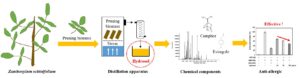
GRAPHICAL ABSTRACT
INTRODUCTION
Zanthoxylum schinifolium, a deciduous spiny shrub, grows abundantly in Korea, China, Taiwan, Manchuria, and Japan (Shin et al. 2008). Evidence shows that this species has been used extensively in traditional Chinese medicine, under the name of huajiao, due to its detoxification properties (Diao et al. 2013). This class of shrub has a unique aromatic flavor in its fruits and leaves, and therefore it has been traditionally used as an edible or medicinal plant. Consequently, this species has attracted attention as a promising medicinal deciduous shrub. The effectiveness of the antibacterial and anticancer activities of some compounds of this species have recently been scientifically confirmed (Choi et al. 2008). The bioactive compounds for medical use can be found in all plant parts of Z. schinifolium (Tuyen et al. 2021). However, the components of the extracts obtained from various parts of the same plant can fluctuate, leading to important compositional dissimilarities. For example, two different essential oils are produced from a cinnamon tree, one from leaves containing 87% eugenol as the major component, and the other from bark containing 97% cinnamaldehyde (Singh et al. 2007). As previously reported, the medical efficacy according to the bioactive compounds of the extract of Z. schinifolium may also differ from part to part (Tuyen et al. 2021).
Previous studies have indicated the presence of coumarins and flavonoids in the extract of Z. schinifolium fruit (Iseli et al. 2007). Essential oils of Z. schinifolium fruit have been used as cosmetic materials to treat allergies and inflammation. In a previous study (Guo et al. 2018), the essential oils obtained from some Z. schinifolium fruits were screened for anti-allergic or anti-inflammatory activities.
Phytochemical studies have indicated the presence of alkaloids, flavonoids, coumarins, triterpenoids, and steroids in the leaves of Z. schinifolium (Cui et al. 2009). Essential phytochemicals of oil obtained from Z. schinifolium leaves exhibit anti-platelet aggregation and anti-microbial, anti-inflammatory, and anti-cancer activities, thus making them ideal alternatives to pure chemical-based compounds (Cui et al. 2009).
However, the parts used in most Z. schinifolium studies are limited to fruits and leaves, and there have been few studies using the branches. In addition, because branches account for 68% of Z. schinifolium, the comprehensive use of the branches of Z. schinifolium is an interesting approach from an economic perspective (Gullón et al. 2018). Z. schinifolium-derived pruning biomass branch comprises different wastes and by-products from Z. schinifolium tree cultivation and the Z. schinifolium fruit oil industry that meet these requirements. Z. schinifolium trees are pruned mainly in the winter between November to January every year, and it is necessary to concentrate the nutrients to increase the fruit yield and facilitate harvesting. This pruned biomass branch does not have industrial applications and is usually burnt or left in the field to serve as a fertilizer. However, both practices lead to environmental risks. Therefore, it is necessary to study the potential of using Z. schinifolium branches as a raw material.
To date, most of the fruits and leaves of Z. schinifolium have been extracted with essential oils, chemical components have been extracted and analyzed, and various pharmacological effects have been studied (Guo et al. 2018; Kim et al. 2018; Tuyen et al. 2021). However, the yield of oil separated from the fruit or leaves of Zanthoxylum is extremely low, at approximately 2% to 7% levels (Paudel et al. 2017). Hydrosol, also called hydrolate or oral water, is a hydrophilic fraction that can be obtained during the essential oil extraction process as a byproduct (Aydin and Caniklioğlu 2021). The hydrosol contains a portion of the essential oil components dissolved in and remaining in the distillation water. It has an extremely pleasant aroma and has been recognized for its commercial applications (Tannous et al. 2004). Unfortunately, limited reports are available on this aspect of Z. schinifolium. To the best of the authors’ knowledge, there have been several reports on the chemical components of the essential oil extracted through conventional methods from Z. schinifolium, but no data concerning the chemical components and anti-allergic inflammatory effect of hydrosol are available in the literature.
The objectives of this study were to analyze the volatile chemical components of hydrosol from Z. schinifolium branches and to provide scientific evidence to justify the anti-allergic inflammatory effect. In the present study, hydrosol was obtained through a hydro-distillation from some Z. schinifolium branches and screened for anti-allergic or anti-inflammatory activities. To the best of the authors’ knowledge, this is the first evidence of the effects of hydrosol extracted from Z. schinifolium branches on allergic inflammation.
EXPERIMENTAL
Materials
Branches of Z. schinifolium were used in this study. The branches were collected in the experimental forest of the National Institute of Forest Science, Jin-ju City, Republic of Korea, in August 2020. The taxonomical identification was established by Dr. Hak Gon Kim at Gyeongsangnam-do Forest Environment Research Institute, and the voucher specimen code applied was WTFRC 10030535.
Methods
Collection of hydrosol from branch of Z. schinifolium
The hydrosol in the fresh branches were extracted by performing steam distillation using a distillation apparatus (Fig. 1). Each branch was cut to a length of 2 cm, and for each load, 25 kg of branches were placed in a distillation vessel. Steam was produced from the boiling water (75 L) at the bottom of the vessel, and the cut branches were distilled at a working temperature of 100 °C for 2 h. The steam with the essential oil was carried to a water-cooled, parallel-piped multi-tubular steel condenser, where it was condensed. The condensed water was hydrated, and the hydrosol was separated in a 10 L Florentine flask. Without performing further purification or filtration, the hydrosol was collected from the apparatus and stored in sealed vials at 4 °C in the dark. The yield was calculated as follows: (dried sample before extraction – dried sample after extraction)/dried sample before extraction ×100 and was found to be 17 %.
 |
| Fig. 1. Distillation apparatus for collection of hydrosol from Z. schinifolium branch |
Gas chromatography-mass spectrometry analysis
The volatile components of hydrosol obtained from the branch of Z. schinifolium was analyzed using gas chromatography-mass spectrometry (GC-MS) (Clarus 600 GC-MS, Perkin Elmer, Shelton, CT, USA). The analysis column used was PerkinElmer Elite-5ms (30 mm × 0.3 mm × 0.25 μm). Helium gas at 1.0 mL/min was used as the mobile phase. The oven temperature was increased from 40 °C to 100 °C at a rate of 10 °C/min and then maintained for 1.0 min. Next the temperature was raised to 230 °C at a rate of 10 °C/min and then maintained for 5 min. The temperature of the injector was set to 200 °C, and the temperature of the detector was set to 250 °C. The analyzed results were identified using the NIST Mass Spectral Search Program (Version 2.0 g, National Institute of Standards and Technology, Gaithersburg, MD, USA).
Cell culture and reagents
The rat cell line RBL-2H3 was supplied by the Korean Cell Line Bank (Seoul, Republic of Korea). The RBL-2H3 cells were cultured in Dulbecco-modified Eagle’s medium (DMEM) (Gibco Labs, Burlington, Ontario, Canada) supplemented with 10% FBS (Bovine serum FBS, Gibco, Rockville, MD, USA) and 1% penicillin-streptomycin in an atmosphere of 5% CO2 in a humidified 37 °C incubator. The RAW264.7 macrophages were purchased from American Type Culture Collection (Manassas, VA, USA) and were cultured in DMEM with 10% FBS and 1% penicillin-streptomycin. Quercetin, 1-chloro-2,4-dinitrobenzene, DNCB, and dexamethasone were purchased from Sigma-Aldrich (St. Louis, MO, USA). 2,4-Dinitrophenylated bovine serum albumin (DNP-BSA), was purchased from Invitrogen (Carlsbad, CA, USA) by Thermo Fisher Scientific. In addition, β-act, SNAP23, Syntaxin4, VAMP7, and VAMP8 were purchased from R&D Systems, Inc. (Minneapolis, MN, USA). Hydrosol was diluted using DMSO to a concentration of 25, 50, 75, and 100 ppm for the working concentration.
Microculture tetrazolium (MTT) assay for cell viability
RBL-2H3 cells were seeded into 96-well plates (SPL Life Sciences Co., Pocheon, Republic of Korea) at 1.5 × 10⁵ cells/mL overnight under an atmosphere of 5% CO2 in a humidified 37 °C incubator, and then treated with hydrosol at a concentration of 25 ppm to 100 ppm for 24 h. Cell proliferation was assayed using tetrazolium dye (MTT solution) (Promega, Madison, WI, USA) according to the manufacturer’s instructions, and absorbance was read using an absorbance microplate reader (SpectraMax 190, Molecular Devices LLC, San Jose, CA, USA) at 490 nm.
SNARE phosphorylation assay
RBL-2H3 cells were pretreated with hydrosol (25 to 100 ppm) for 8 h at 37 °C in a CO2 incubator. The RBL-2H3 cells (1 × 10⁵) were lysed in 1 mL of lysis buffer containing a protease inhibitor cocktail (Calbiochem, Germany) (Hiramatsu et al. 2010). Lysates were centrifuged at 13,000 rpm at 4 °C to remove cell debris and pretreated for 2 h at 4 °C with 100 μL of protein. Antibodies to a SNARE protein (SNAP23, Syntaxin4, VAMP2, and VAMP8) were added to each lysate and allowed to bind at 4 °C for 2 h. After adding protein G suspension to the antibody-antigen complexes, mixtures were allowed to settle overnight. After vigorously washing with lysis buffer, precipitates were subjected to immunoblot analysis. Whole cell lysates and immune-precipitated proteins were separated on 12% SDS-PAGE gels and transferred to nitrocellulose membranes (Whatman, Maidstone, UK) for analysis. Membranes were blocked at room temperature for 1 h in Tris-buffered saline with 0.1% Tween® 20 detergent (TBST) containing 5% powdered skim milk and 0.5% BSA, then probed with β-actin (1: 1000), SNAP23 (1:1000), Syntaxin4 (1:2000), VAMP7 (1:1000), or VAMP8 (1:1000) primary antibodies overnight. After washing with TBST, the membranes were treated with a secondary antibody (anti-mouse IgG; A4416, Sigma) coupled with horseradish peroxidase. Signal was detected using enhanced chemi-luminescence (ECL) solution.
β-hexosaminidase release by RBL-2H3
β-Hexosaminidase is a marker of mast cell degranulation (Nagai et al. 2012). The RBL-2H3 cells (2 × 10⁵ cells/mL) were seeded in 500 μL/well of culture medium into 24-well plates and cultured overnight. Cells were washed with HEPES-buffered, and an anti-dinitrophenyl (DNP)-antibody. After incubation in IgE (100 ng/mL, Sigma) for 3 h, cells were incubated for 30 min with vehicle, hydrosol (25 to 100 ppm), or quercetin (positive control; 20 µM). Cells were then stimulated with a DNP-group conjugated to bovine serum albumin (BSA) to initiate antigen-induced degranulation (200 ng/mL, DNP-BSA). Degranulation was quantified by measuring β-hexosaminidase activity in supernatants for 30 min at 37 °C in 4-(2-hydroxyethyl)-1-piperazineethanesulfonic acid (HEPES)-buffered saline, using a colorimetric biochemical assay. Supernatants were transferred to 96 well plates (50 μL/well), and 50 μL of substrate solution was added. Plates were incubated at 37 °C for 3 h. After stopping reactions in 100 μL Na2CO3-NaHCO3 buffer, the absorbance was measured at 405 nm using a microplate reader (SpectraMax 190, Molecular Devices LLC, San Jose, CA, USA). Secretory activity was calculated as the relative total β-hexosaminidase content in the cells. Total β-hexosaminidase content was determined using cells lysed with 0.1% Triton X-100.
ROS generation measurement
To measure the intracellular reactive oxygen species (ROS), measurements were conducted using 27′-dichloro-fluorescein diacetate (DCF-DA, Sigma-Aldrich, St. Louis, MO, USA), which emits fluorescence when reacting with free radicals. After incubating RAW 264.7 cells in a 96-well plate at a concentration of 5 × 10⁴ cells/well, the cells were treated with hydrosol at 25 to 100 ppm or a positive control Bay 11-7082 (20 µM) for 4 h and then treated with 1 µg/mL lipopolysaccharide (LPS) for 24 h; the medium was then fully removed. Then, DCF-DA was added to PBS and reacted at 37 °C for 30 min, and the active oxygen was then confirmed using an absorbance microplate reader (SpectraMax 190, Molecular Devices LLC, San Jose, CA, USA) at an excitation wavelength of 485 nm and an emission wavelength of 530 nm.
Nitric oxide analysis
The concentration of NO was estimated indirectly by measuring the nitrite concentration in the culture medium using Griess Reagent. The RAW 264.7 cells were pretreated with hydrosol (25 to 100 ppm) or Bay 11-7082 (positive control; 20 µM). After 1 h, 100 ng/mL of LPS was added, and cells were incubated for 18 h. Griess Reagent was added in 1:1 ratio to 50 μL/well of conditioned culture media. Reactions were incubated at room temperature for 10 min. The absorbance was measured at 570 nm using an absorbance microplate reader (SpectraMax 190, Molecular Devices LLC, San Jose, CA, USA).
Statistical analyses
All experiments were repeated at least three times, and data were obtained in triplicate. The statistical significance of the differences between the control and experimental results was estimated through a t-test using R-project (version 2.6.1, R Foundation, Auckland, New Zealand).
RESULTS
Chemical Components of Hydrosol
The chemical components of the hydrosol obtained from the Z. schinifolium branch are listed in Table 1, and five main components were identified. Estragole was the predominant component, accounting for 50.86% of the total. This was followed by camphor (30.68%), R(+)-limonen (3.75%), elemol (1.89%), and linalool (1.10%) (Table 1). A gas chromatogram of the hydrosol is presented in Fig. 2. In previous studies, the essential oil content of Z. schinifolium fruit was reported by Iseli et al. (2007) as limonene (21.63%), 4-terpineol (14.50%), and α-terpineol acetate (5.33%).

 |
| Fig. 2. Gas chromatogram of hydrosol obtained from Z. schinifolium branch; Identification of the major peaks: 1: Linalool; 2: Camphor; 3: Estragole; 4: R(+)-Limonen; 5: Elemol |
Tuyen et al. (2021) reported that the major components of essential leaf oil from Z. schinifolium were limonene (44.3%), β-caryophyllene (12.5%), linalool (11.0%), germacrene D (5.3%), and α-pinene (4.9%), and the major components of the essential fruit oil were n-pentadecane (34.8%), sabinene (18.3%), and n-heptadecane (4.7%), and the major components of the essential Z. schinifolium branch oil were 2-undecanone (72.3%), β-caryophyllene (5.8%), and germacrene D (4.0%). As reported in above studies the volatile components may vary depending on the parts of the same species of plants, which is also shown by the results of this study (Table 2).

Effects of Hydrosol on Cell Viability
The effect of hydrosol on the cell viability was evaluated using an MTT assay in RBL-2H3 cells to ensure that its anti-allergic inflammatory effects were not due to cell death under each condition (Fig. 3). Treatment with hydrosol for 24 h at concentrations of 25 to 100 ppm showed no significant cytotoxic effects. In addition, the toxicity of DMSO used for controlling the concentration of the hydrosol was evaluated, and DMSO showed no toxicity in the RBL-2H3 cells.

Fig. 3. Effect of hydrosol on cell viability in RBL-2H3 cells. The cell viability was tested using MTT assays. The data are representative of three independent experiments and expressed as the mean ± standard error of the mean (SEM
Inhibition of Phosphorylation of SNAREs by Hydrosol
The inhibition of the phosphorylation of SNAREs is a key indicator of reduced allergic symptoms (Hoesel and Schmid 2013). Therefore, the effects of hydrosol on the phosphorylation of SNAREs (Fig. 4) were examined. In mast cells, similar to other secretory cells such as neuronal cells and pancreatic β cells, SNARE proteins are involved in membrane fusion between secretory granules and the plasma membrane following a Ca2+ increase and protein kinase C (PKC) activation. The major SNAREs involved in mast cell exocytosis are SNAP23, Syntaxin4, VAMP7, and VAMP8 (Morita et al. 2011). As shown in Fig. 4, the hydrosol inhibited the phosphorylation of all four SNAREs, with remarkable inhibitory effects on Syntaxin4 and VAMP7. SNAP23 and VAMP8 were inhibited at 75 ppm hydrosol, and Syntaxin4 and VAMP7 were inhibited at 25 ppm and 37.5 ppm hydrosol respectively. In particular, the inhibition of Syntaxin4 was confirmed at 25 ppm, which is the lowest concentration of hydrosol. These results demonstrated that hydrosol was effective in inhibiting the phosphorylation of SNAREs to reduce allergic symptoms.
 |
| Fig. 4. Effects of hydrosol on phosphorylation of SNAREs; effects of hydrosol on phosphorylation of SNAP23, Syntaxin4, VAMP7, and VAMP8 revealed by Western blotting
|
 |
| Fig. 5. Effect of hydrosol on β-hexosaminidase secretion in RBL-2H3 cells; experiments were conducted in quadruplicate and expressed as mean ± SEM; +++: p < 0.001 compared DNP-IgE; *: p < 0.05 compared DNP-BSA; ***: p < 0.001 compared DNP-BSA |
Effect of Hydrosol on β-hexosaminidase Secretion in RBL-2H3 Cells
The release of β-hexosaminidase is proportional to the extent of degranulation and is considered a biomarker for mast cell degranulation (Huang et al. 2015; Zeng et al. 2016). The hydrosol was also tested for β-hexosaminidase activity in order to investigate whether its effects were due to the inhibition of enzyme activity or degranulation. Treatment with hydrosol significantly suppressed the β-hexosaminidase activity in the DNP-BSA-stimulated RBL-2H3 cells. As a result, 50 and 100 ppm hydrosol were inactive against the enzyme activity of β-hexosaminidase. In addition, 100 ppm hydrosol in DNP-BSA-stimulated RBL-2H3 cells inhibited β-hexosaminidase release to a similar extent as quercetin, indicating that hydrosol may be a promising new anti-allergic agent (Fig. 5).
Effect of Hydrosol on Production of Inflammatory Mediators in LPS-Activated RAW264.7 Cells
Pre-treatment with hydrosol significantly inhibited ROS, achieving 50 to 100 ppm, which was significantly different from the control group (Fig. 6A).

Fig. 6. Effect of hydrosol on the production of inflammatory mediators in LPS-activated RAW264.7 cells: RAW264.7 cells seeded into a 48-well plate were pretreated with hydrosol (25 to 100 ppm) or Bay 11-7082 (positive control, 20 µM) for 2 h prior to the addition of LPS (500 ng/mL) for 24 h. ROS (A) in the supernatants was measured using ELISA; and NO production (B) in the supernatants was detected through a Griess reagent. The data are representative of three independent experiments and expressed as mean ± SEM (∗P < 0.05; ∗∗∗P < 0.001 versus with Bay 11-7082).
The suppressive effects of 100 ppm hydrosol approached the effect of the reference compound Bay 11-7082 at 20 µM. In addition, the NO production was significantly decreased by hydrosol at a concentration of 25 ppm, which implied anti-inflammatory activity in LPS-induced RAW264.7 macrophages (Fig. 6B). It was confirmed that hydrosol inhibited ROS and NO production in a concentration-dependent manner. In particular, 100 ppm hydrosol suppressed ROS as well as NO, similar to the positive control. Therefore, it was concluded that 100 ppm hydrosol is effective in inhibiting ROS and NO that mediate inflammation.
Inhibition of Phosphorylation of SNAREs by Main Components of Hydrosol
The main components of the hydrosol were estragole (50.86%) and camphor (30.68%). Camphor, the main component of hydrosol, inhibited the phosphorylation of Syntaxin4, VAMP7, and VAMP8 SNAREs (Fig. 7). It can therefore be indirectly confirmed that the anti-inflammatory activity of hydrazole is induced through camphor compound of the main components.
 |
| Fig. 7. Effects of main components on phosphorylation of SNAREs; effects of hydrosol on phosphorylation of SNAP23, Syntaxin4, VAMP7, and VAMP8 as revealed through Western blotting |
CONCLUSIONS
1. In the present study, the hydrosol extracted from a branch of Z. schinifolium was screened for anti-allergic and anti-inflammatory activities, which showed a high inhibitory activity on the mast cell degranulation.
2. About five main components from hydrosol were identified, and estragole was the predominant component (50.86%), followed by camphor (30.68%).
3. The present study revealed that hydrosol inhibits degranulation in DNP-BSA-stimulated mast cells, and is an effective inhibitor of LPS-induced pro-inflammatory mediators in RAW264.7 macrophages by suppressing ROS and NO.
4. These findings suggest that hydrosol obtained from the branch of Z. schinifolium may serve as a potential therapeutic candidate for the treatment of anti-allergic and anti-inflammatory diseases.
ACKNOWLEDGMENTS
This study was carried out with the financial support of ‘R&D Program for Forest Science Technology (Project No. “2020186D10-2122-AA02”)’ provided by Korea Forest Service (Korea Forestry Promotion Institute).
REFERENCES CITED
Aydin, S., and Caniklioğlu, A. (2021). “Antimicrobial and antioxidant potentials, total phenolic contents of some herbal waters,” Eur. J. Biol. Res. 11(2), 203-211. DOI: 10.5281/zenodo.4501542
Choi, H. J., Song, J. H., Kwon, D. H., Baek, S. H., and Ahn, Y. J. (2008). “Antiviral activity of Zanthoxylum species against Influenza virus,” Korean J. Med. Crop Sci. 16, 273-278.
Cui, H. Z., Choi, H. R., Choi, D. H., Cho, K. W., Kang, D. G., and Lee, H. S. (2009). “Aqueous extract of Zanthoxylum schinifolium elicits contractile and secretory responses via β1-adrenoceptor activation in beating rabbit atria,” J. Ethnopharmacol 126(2), 300-307. DOI: 10.1016/j.jep.2009.08.025
Diao, W. R., Hu, Q. P., Feng, S. S., Li, W. Q., and Xu, J. G. (2013). “Chemical composition and antibacterial activity of the essential oil from green huajiao (Zanthoxylum schinifolium) against selected foodborne pathogens,” J. Agric. Food Chem. 61(25), 6044-6049. DOI: 10.1021/jf4007856
Gullón, P., Gullón, B., Dávila, I., Labidi, J., and González-Garcia, S. (2018). “Comparative environmental life cycle assessment of integral revalorization of vine shoots from a biorefinery perspective,” Sci. Total Environ 624, 225-240. DOI: 10.1016/j.scitotenv.2017.12.036
Guo, R. H., Park, J. U., Jo, S. J., Ahn, J. H., Park, J. H., Yang, J. Y., Lee, S. S., Park, M. J., and Kim, Y. R. (2018). “Anti-allergic inflammatory effects of the essential oil from fruits of Zanthoxylum coreanum Nakai,” Front. Pharmacol. 9, 1441-1453. DOI: 10.3389/fphar.2018.01441
Hiramatsu, H., Tadokoro, S., and Hirashima, N. (2010). “Latrotoxin induces degranulation in RBL2H3 cells transfected with latrophilin,” Toxicon 56, 1372-1380. DOI: 10.1016/j.toxicon.2010.08.002
Hoesel, B., and Schmid, J. A. (2013). “The complexity of NF-κB signaling in inflammation and cancer,” Molecular Cancer 12(1), 1-15. DOI: 10.1186/1476-4598-12-86
Huang, L., Li, T., Zhou, H., Qiu, P., Wu, J., and Liu, L. (2015). “Sinomenine potentiates degranulation of RBL-2H3 basophils via up-regulation of phospholipase A2 phosphorylation by Annexin A1 cleavage and ERK phosphorylation without influencing on calcium mobilization,” Int. Immunopharmacol. 28(2), 945-951. DOI: 10.1016/j.intimp.2015.04.029
Iseli, V., Potterat, O., Hagmann, L., Egli, J., and Hamburger, M. (2007). “Characterization of the pungent principles and the essential oil of Zanthoxylum schinifolium pericarp,” Pharmazie 62, 396-400. DOI: 10.1691/ph.2007.5.6701
Kim, Y. N., Park, M. J., Zheng, Z., Lee, S. S., Yoo, K. H., and Kim, H. (2018). “Inhibitory effects of essential oil from Zanthoxylum coreanum Nakai on melanin production: Protection against re-pigmentation after laser treatment,” Medica Lasers; Engineering, Basic Research, and Clinical Application 7(2), 62-68. DOI: 10.25289/ml.2018.7.2.62
Morita, Y., Tadokoro, S., Sasai, M., Kitamoto, D., and Hirashima, N. (2011). “Biosurfactant mannosyl-erythritol lipid inhibits secretion of inflammatory mediators from RBL-2H3 cells,” Biochimica et Biophysica Acta (BBA)-General Subjects 1810(12), 1302-1308. DOI: 10.1016/j.bbagen.2011.07.002
Nagai, K., Fukushima, T., Oike, H., and Kobori, M. (2012). “High glucose increases the expression of proinflammatory cytokines and secretion of TNFα and β-hexosaminidase in human mast cells,” Eur. J. Pharmacol. 687(1-3), 39-45. DOI: 10.1016/j.ejphar.2012.04.038
Paudel, K., Bhatt, T. D., Adhikari, A. K., and Basyal, C. (2017). “Composition comparison of essential oils of Zanthoxylum armatum DC by GC-MS,” J. Plant Resour. 15(1), 81-85.
Shin, H. T., Lim, J. T., and Goo, K. H. (2008). “Selection of sancho (Zanthoxylum schinifolium S. et Z.) cultivar for production of essential oils,” J. Korean Nat. 1(2), 215-218. DOI: 10.1016/S1976-8648(14)60139-0
Singh, G., Maurya, S., De Lampasona, M. P., and Catalan, C. A. (2007). “A comparison of chemical, antioxidant, and antimicrobial studies of cinnamon leaf and bark volatile oils, oleoresins, and their constituents,” Food Chem. Toxicol. 45(9), 1650-1661. DOI: 10.1016/j.fct.2007.02.031
Tannous, P., Juliani, R., Wang, M., and Simon, J. (2004). “Water balance in hydrosol production via steam distillation: Case study using lavandin (Lavandula x intermadia),” in: New Use Agriculture and Natural Plant Products and ASNAPP Program, New Brunswick, Newark and Camden, NJ, USA, pp. 1-15.
Tuyen, T. T., Quan, P. M., Thu Le, V. T., Toan, T. Q., Nghi, D. H., Bach, P. C., Inh, C. T., Hanh, N. P., Vien, T. A., Hong Minh, P. T., Long, et al. (2021). “Chemical composition, antimicrobial, and cytotoxic activities of leaf, fruit, and branch essential oils obtained from Zanthoxylum nitidum grown in Vietnam,” Nat. Prod. Commun 16(1), 1-7. DOI: 10.1177/1934578X20985649
Zeng, H. R., Wang, B., Zhao, Z., Zhang, Q., Liang, M. Y., Yao, Y. Q., Bian, K., and Zhang, W. R. (2016). “Effects of Viola yedoensis Makino anti-itching compound on degranulation and cytokine generation in RBL-2H3 mast cells,” J. Ethnopharmacol 189, 132-138. DOI: 10.1016/j.jep.2016.05.030
Article submitted: April 5, 2021; Peer review completed: June 7, 2021; Revised version received and accepted: June 25, 2021; Published: July 1, 2021.
DOI: 10.15376/biores.16.3.5721-5732