
Abstract
Garlic (Allium sativum L.) being infected by mycotoxigenic fungi is one of the primary factors limiting its nutritional and medical value. Therefore, there is an urgent need to repress mycotoxigenic fungi utilizing safer treatments, possibly involving nanoparticles. Fusarium incarnatum was isolated from garlic (A. sativum L.) that showed fungal contamination and their identification was confirmed using amplified and sequenced internal transcribed spacer nuclear ribosomal DNA regions, which confirmed the isolation of F. incarnatum from all cultivars. Copper oxide nanoparticles (CuONPs) at different concentrations showed inhibitory activity against F. incarnatum growth and mycotoxins, particularly at 400 ppm. The production of F. incarnatum mycotoxins, i.e., beauvericins, fusarins, moniliformin, and enniatins, was inhibited to 62.8%, 45.4%, 58.1%, and 55.0%, respectively at 400 ppm of CuONPs compared to the control. Shrinkage of the F. incarnatum cell membrane and collapsing of the cell walls were recorded via transmission electron microscopy at 400 ppm, but negligible distortion appeared at 100 ppm of CuONPs. CuONPs at 100 ppm encouraged the activity of CMC-ase, zylanase, and amylase, while 200 and 400 ppm promoted less enzyme activity. The current findings suggest that CuONPs have a fungistatic effect on F. incarnatum and their mycotoxins.
Download PDF
Full Article
Copper Oxide Nanoparticles as Fungistat to Inhibit Mycotoxins and Hydrolytic Enzyme Production by Fusarium incarnatum Isolated from Garlic Biomass
Aisha M. H. Al-Rajhi,a Reham Yahya,b,c Mohamed M. Alawlaqi,d Mohamed A. Fareid,e,f Basma H. Amin,g and Tarek M. Abdelghani e,*
Garlic (Allium sativum L.) being infected by mycotoxigenic fungi is one of the primary factors limiting its nutritional and medical value. Therefore, there is an urgent need to repress mycotoxigenic fungi utilizing safer treatments, possibly involving nanoparticles. Fusarium incarnatum was isolated from garlic (A. sativum L.) that showed fungal contamination and their identification was confirmed using amplified and sequenced internal transcribed spacer nuclear ribosomal DNA regions, which confirmed the isolation of F. incarnatum from all cultivars. Copper oxide nanoparticles (CuONPs) at different concentrations showed inhibitory activity against F. incarnatum growth and mycotoxins, particularly at 400 ppm. The production of F. incarnatum mycotoxins, i.e., beauvericins, fusarins, moniliformin, and enniatins, was inhibited to 62.8%, 45.4%, 58.1%, and 55.0%, respectively at 400 ppm of CuONPs compared to the control. Shrinkage of the F. incarnatum cell membrane and collapsing of the cell walls were recorded via transmission electron microscopy at 400 ppm, but negligible distortion appeared at 100 ppm of CuONPs. CuONPs at 100 ppm encouraged the activity of CMC-ase, zylanase, and amylase, while 200 and 400 ppm promoted less enzyme activity. The current findings suggest that CuONPs have a fungistatic effect on F. incarnatum and their mycotoxins.
DOI: 10.15376/biores.17.2.3042-3056
Keywords: Fusarium incarnatum; Garlic; Hydrolytic enzymes; Mycotoxins; Nanoparticles
Contact information: a: Department of Biology, College of Science, Princess Nourah bint Abdulrahman University P.O. Box 84428, Riyadh 11671, Saudi Arabia; b: Medical Microbiology, College of Science and Health Professions, King Saud Bin Abdulaziz University for Health Sciences, Riyadh 14611 Saudi Arabia; c: King Abduallah International Medical Research Center, Riyadh 21423 Saudi Arabia; d: Biology Department, Faculty of Science, Jazan University, Jazan 45142 Saudi Arabia; e: Botany and Microbiology Department, Faculty of Science, Al-Azhar University, Nasr City, Cairo 11725, Egypt.; f:Basic Science Department, Prep Year Deanship, University of Haʼil, Saudi Kingdom.; g: The Regional Centre for Mycology and Biotechnology (RCMB), Al-Azhar University, Cairo, Egypt;
* Corresponding author: tabdelghany.201@azhar.edu.eg; tabdelghany@yahoo.com
GRAPHICAL ABSTRACT
INTRODUCTION
Numerous fungi usually attack cereal grains, seeds, vegetables, fruits, as well as their foodstuff products either in the field or storage places. The ability of these fungi to produce mycotoxins has been documented under particular conditions, which minimizes the marketable and dietary value of these products, as well as results in undesirable health impacts on consumers including humans and animals (Abdelghany 2006). Many scientific papers have reported that Fusarium is considered one of the greatest economically detrimental fungal genera for many reasons, i.e., its phytopathogenic activity, potent producer for mycotoxins, and illness impacts on humans and livestock (Abdelghany 2014; Villani et al. 2019; Agriopoulou et al. 2020; Nofal et al. 2021a). Multiple documents have reported that Fusarium species are responsible for the production of several mycotoxins, e.g., fumonisins, trichothecenes, zearalenone, enniatins, and fusaproliferin (Munkvold 2017; Jajić et al. 2019; Gautier et al. 2020). Recently, Choi et al. (2021) reported the presence of Fusarium spp. in millet samples with the highest frequency of mycotoxin being F. asiaticum, followed by F. acuminatum, F. graminearum, F. incarnatum, F. equiseti, and F. tricinctum besides other species of Fusarium. Occurrences of zearalenone, deoxynivalenol, fumnasine, and nivalenol in these millet samples were also recognized. The trichothecenes A and ZEN found in corn were F. incarnatum (Munkvold 2017). Yazid et al. (2021) showed that three isolates of F. incarnatum produced ZEN, while HT-2 and T-2 toxins were produced by one isolate.
Previously, different strains were isolated from Tunisian sorghum, and the presence of ZEA in sorghum samples varied according to the strain (Lahouar et al. 2017). Villani et al. (2019) mentioned that F. incarnatum–equiseti species complex (FIESC) covers 33 phylogenetically diverse species that have been isolated from diverse biological samples as well as agricultural soils. According to Villani et al. (2019), although miscellaneous mycotoxins were produced by FIESC, its potential to cause mycotoxin contamination in grains and other food products is unclear because the diversity of these species has only been recently documented.
Outside of the nutritional value, the therapeutic potential of garlic (Allium sativum L.) has been previously confirmed and is still the focus of studies, due to the presence of multiple bioactive molecules (Singh and Sahu 2020). Garlic is considered one of the oldest and most imported crops in all countries; therefore its worldwide production has been regularly increasing over the last two decades. Although numerous scientific reports mention the antifungal activity of garlic extract in vitro and in vivo, this activity is quite a paradox, since the garlic plant or its fruits can be colonized by numerous fungi, particularly Fusarium species comprising of F. equiseti, F. proliferatum, F. oxysporum, F. acuminatum, and F. solani (Stankovic et al. 2007; Ignjatov et al. 2018; Miriam et al. 2021; Paul et al. 2021). The deteriorative efficacy of Fusarium spp. as well as other fungi on plants are due to the secretion of cell wall-degrading enzymes (CWDEs), e.g., lignocellulases, lipases, and proteases, which are responsible for the damage of the cell wall of plants (Nofal et al. 2021b; Al-Rajhi et al. 2022). Furthermore, the produced fungal toxins suppress the immune system of the plant, which helps the fungi successfully attack the plant (Lakshmipriya et al. 2020). Mallebrera et al. (2018) mentioned that Fusarium toxins enniatins and beauvericins encourage DNA fragmentation in addition to apoptosis through disorder in the mitochondrial pathways.
The search for effective approaches to eliminate pathogenic fungi and their toxins has become a global demand, so scientists turned to applying nanotechnology as a promising and potentially safe method for controlling fungal proliferation. Nanoparticles (NPs) have been applied as fungicide to restrict growth and mycotoxin production (Abdelghany et al. 2018a; Ganash et al. 2018; Jesmin and Chanda 2020). However, the activity of NPs on the production of mycotoxins has not been researched enough yet. More recent attention has been focused on the application of copper nanoparticles (CuNPs) and copper oxide nanoparticles (CuONPs) in these fields (Khamis et al. 2017; Abdelghany et al. 2020). It has been found that CuONPs suppresses Fusarium culmorum, F. graminearum, F. oxysporum, F. solani, Penicillium chrysogenum, Aspergillus flavus, and Alternaria alternate, and Aspergillus niger growth (Ghasemian et al. 2012; Shende et al. 2015; Abdelghany et al. 2020). The effect of CuONPs exhibited genotoxicity in F. solani and Penicillium digitatum as a result of exposure to CuONPs (Khamis et al. 2017). The repression of mycotoxigenic fungi growth is an area of interest for numerous investigators; therefore, the current study aimed to evaluate the activity of CuONPs against the growth, ultrastructure, and mycotoxins, as well as hydrolytic enzyme production of F. incarnatum isolated from garlic (Allium sativum L.).
EXPERIMENTAL
Isolation and Identification of Mycotoxins Producing Fungus
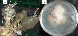
Cloves of garlic (Allium sativum L.) that showed fungal infection (as shown in Fig. 1A) from a field in Menofia Governorate, Egypt were separated, peeled off, and the surface was disinfected using 5% NaOCl for 2 min. They were rinsed in sterile water using three successive baths of for 1 min each, then dried using sterile filter paper to remove the excess water. Small, infected tissue samples with symptoms of fungal appearance (1 cm in length) were plated onto potato dextrose agar medium (PDA) amended with streptomycin sulphate (3%) and incubated for 6 d at a temperature of 28 °C until fungal growth appeared. The hyphal tips of the appeared Fusarium-like colonies were purified by being transferred to PDA plates and were incubated at a temperature of 28 °C until the purified fungus appeared, which was kept for further study (Dhingra and Sinclair 1995). The purified fungus was morphologically identified by the presence of macroconidia, microconidia, chlamydospores, as well as by the observation of the growth rate and the color of the colony (Nelson et al. 1983; Leslie and Summerell 2006).
Molecular Identification of the Pathogen
The purified and morphologically identified fungus was subjected to molecular identification (Abdelghany et al. 2018b; Hami et al. 2021). For identification, fungal DNA was extracted using a Microprep Kit of quick-DNA fungi/bacteria (Zymo research; D6007) according to the procedure outlined by Sigma Scientific Services Company. Maxima Hot Start PCR Master Mix (Thermo; K1051) was utilized for the PCR. Amplification of the DNA was done using forward primer ITS1-F (5′- TCCGTAGGTGAACCTGCGG-3′) and reverse primer ITS4-R (5′- TCCTCCGCTTATTGATATGC-3′). Various thermal cycler conditions throughout the PCR protocol were used, starting with 1 cycle at a temperature of 94 °C for 6 min, which was required for the initial denaturation, followed by 35 cycles for 45 s at a temperature of 94 °C, which was required for another denaturation, then 35 cycles for 45 s at a temperature of 56 °C, which was required for annealing, and ending with 35 cycles for 60s at a temperature of 72 °C, which was required for extension. The fungus DNA was scanned via gel electrophoresis. Sequences homologous at NCBI (http://ncbi.nlm.nih. gov/BLAST) were analysis via BLAST. The neighbor joining manner was used for the reconstruction of the evolutionary tree.
Copper oxide Nanoparticles and Rizolex-T50
Copper oxide nanoparticles (CuONPs) were purchased from Nawah Scientific, Cairo, Egypt. Particles were less than 100 nm and came in powder form. The size was confirmed by scanning electron microscopy (FEI Company, Hillsboro, Oregon-USA) at EDRC, DRC, Cairo). Rizolex-T 50 (20% Tolclophos-methyl and 30% Thiram) was applied as a chemical fungicide, which was purchased from Sumitomo Chemical, Tokyo, Japan.
Assessment of Copper Oxide Nanoparticles (CuONPs) Activity on Growth and Ultra-Structures of Pathogen
The inhibitory action of CuONPs at different concentrations against F. incranatum growth was achieved using Petri dishes containing Czapek agar media. Rizolex-T50 was applied as a positive control for fungal growth. The plates were centrally point-inoculated with a 0.5 mm disc from 5-day-old colony cultures of the pathogen, and then incubated for 7 d at a temperature of 28 °C. At the end of the incubation period, the radius of the colony was measured to calculate the inhibition %, according to Eq. 1,
 (1)
(1)
where CRC (mm) is the colony radius of the control and CRT (mm) is the colony radius of the treatment (Note: CuONPs nil medium was used as the control).
The tips of the growing fungus hypha at different concentration of CuONPs were cut and fixed using 5% gluteraldehyde for 1 d. Then, the samples were washed in phosphate buffer (pH 7.2) 3 times, followed by the elimination of the excess buffer. Next, the segments were rinsed for 2 h in osmium tetraoxide diluted up to 1%, followed by the removal of osmium tetraoxide. The prepared segments were dehydrated by a sequence of different levels of ethanol, ranging from 50% to 95%. The segments were dried with absolute ethanol, and then dipped in propylene oxide for 1 h. The segments of hyphae were placed in propylene oxide and Epon 812 resin at a 2 to 1 ratio, followed by being placed in pure resin for 12 h, before finally being placed in an oven for 48 h at a temperature of 60 °C . Blocks (50 nm) were segmented via ultra-microtome, then stained with uranyl acetate-lead citrate 500A and examined at the Regional Center for Mycology and Biotechnology, Al-Azhar University, Cairo, Egypt via transmission electron microscopy (TEM) (C Joel Jem- 1200 EX II. Acc. Voltage 120 KV. MAG- medium) (Abdelghany et al. 2021).
Assessing Copper Oxide Nanoparticles (CuONPs) Activity on Mycotoxins Production
Sterilized potato dextrose broth medium supplemented with different concentrations of CuONPs (50 mL in a 250 mL conical flask) was inoculated with F. incranatum and then incubated for 10 d at a temperature of 28 °C. At end of the incubation period, the fungal mycelia were decanted, and the filtrate medium was extracted with 50 mL of chloroform and methanol (at a 2 to 1 ratio), followed by desiccation to concentrate the extracted solvent (El-Taher et al. 2012). Then the extract was subjected to LC1620A liquid chromatography for mycotoxin analysis. The inject volume was 20 uL, the mobile phase water to acetonitrile to methanol was 5 to 4 to 1, the flow was 1.540 mL/min, and the current detector UV-Detector was at a wavelength of 254 nm. The mycotoxins were identified by comparing the retention time of the analyte with labeled internal mycotoxin standards.
Assessment of the Copper Oxide Nanoparticles (CuONPs) Activity on Hydrolytic Enzyme Production
Three substrates, including 0.5% carboxymethyl cellulose, 0.5% xylan, and 2% starch (Sigma-Aldrich, St. Louis, MO), were used for stimulating the production of CMC-ase, xylanase, and amylase, respectively (El-Katatny 2010). The sterilized minimal synthetic medium (MSM) (NH4NO3 1.0 g/L, K2HPO4 0.9 g/L, KCl 0.2 g/L, MgSO4·7H2O 0.2 g/L, MnSO4 0.002 g/L, FeSO4·7H2O 0.002 g/L, ZnSO4 0.002 g/L, and agar 20 g/L) containing the appropriate substrate and different concentrations of CuONPs (at a pH of 5.5 adjusted by 50 mM acetate buffer) in a plate was inoculated with the isolated fungus in the center of plate and incubated for 5 d. At the end, the enzymes activities were visualized via staining the agar plates with 0.2% Congo red dye for 15 min, followed by washing with a 1 M NaCl solution for detecting the CMC-ase and xylanase (Herculano et al. 2011). The amylase was detected by flooding the plate with 1% iodine from 2% potassium iodide. The enzymatic index (EI) was determined by measuring the enzymatic halo (diameter of colony with the clear zone) according to Eq. 2,
 (2)
(2)
Statistical Analysis
All the results were analyzed via SPSS software (version 14, IBM, Armonk, NY). Calculations were done for the mean ± SD (standard deviation) of three replicates.
RESULTS AND DISCUSSION
Fungal Identification of the Infected Cloves of Garlic Biomass
The collected samples of garlic were covered by a white mycelium (as shown in Fig.1A), with soft and spongy bulbs.

Fig. 1. Garlic samples infected with F. incarnatum (A); colony of F. incarnatum on a Petri dish containing PDA medium (B); and the phylogenetic tree of the F. incarnatum strain JL5-2 (C)
The isolated fungus was morphologically identified as F. incarnatum, where at the initial stage the aerial mycelia were white (Fig. 1B) and turned to off-white, pale pink, and grey in the later phases, in addition to the microscopic examination. Molecular characterization of the isolate was conducted to recognize and confirm that the identity isolate was closely related to F. incarnatum. The phylogenetic tree demonstrated the similarity of the isolate to F. incarnatum (Fig. 1C). A BLAST search in the NCBI database via ITS sequences (Fig. 2) showed 99% to 100% similarities with F. incarnatum. F.incarnatum has been isolated from various plant material in different countries as well as other species, as mentioned in numerous reports. For example, it was detected on rapeseed in Iran (Nemati et al. 2015). Other Fusarium spp. were isolated from garlic, e.g., F. proliferatum (Mondani et al. 2021). Recently, F. proliferatum, F. solani, F. acuminatum, F. oxysporum, F. redolens, and F. subglutinans have been detected in rot garlic (A. sativum) (Gálvez and Palmero 2021). Previously, Penicillium, Aspergillus, Botrytis, and Fusarium genera were detected in garlic bulbs in Egypt, but Fusarium spp. represented the most prevalent genera (Moharam et al. 2013).

Fig. 2. The ITS sequences of the closely related fungal strains retrieved from the NCBI GenBank database and the cluster analysis of F. incarnatum
Inhibitory Action of Copper Oxide Nanoparticles (CuONPs) Against Fungus Growth
The inhibitory action of CuONPs (size less than 100 nm shown in Fig. 3) in terms of F. incarnatum growth and sporogenesis was observed to be dependent on the concentration (as shown in Table 1). The obtained results indicated that the inhibition of growth reached up to 81.70% at 400 ppm when compared to the control zero %. According to the microscopic analysis and spore counting, sporulation (%) decreasing was recorded as the CuONPs concentration increased. Although the inhibitory action of the chemical fungicide (Rizolex-T50) and 400 ppm of CuONPs was approximately in a similar narrow spectrum (85.36 % and 81.70%, respectively), but the sporulation (%) was far apart from each other (10 and 29%, respectively).

Fig. 3. Scanning electron micrograph of CuONPs. Magnification 12000X
Table 1. Colony Diameter, Inhibition and Sporulation of F. incarnatum at Various Copper Oxide Nanoparticles (CuONPs) Concentrations

Recent results showed the substantial suppression of F. oxysporum spore propagation after exposure to CuONPs (Ashraf et al. 2021). The inhibitory activity of CuONPs and chemical fungicide on fungal growth was confirmed and evaluated also in broth medium, which indicated that growth reduced with increasing CuONPs concentrations (Fig. 4).
The fungicidal activity of CuONPs was reported against multiple fungi. For instance, Fusarium solani and Penicillium digitatum had full inhibition at 60 µg/mL and 20 µg/mL, respectively (Khamis et al. 2017). In another study, the controlling effect of CuONPs was recorded for Penicillium expansum, Alternaria alternata, and A. solani (Nemati et al. 2015).

Fig. 4. Growth of F. incarnatum at different concentrations of CuONPs (100, 200, and 400 ppm) in broth medium
A mechanistic study of NPs against various fungi has been investigated. For instance, injury of the lipid bilayer in fungal cell membranes, as well as the formation of unusual swells on the surface of the hyphae, which led to hyphae deformation, were observed as a result of exposure to ZnO nanoparticles (He et al. 2011).
In the current research, the TEM showed that CuONPs caused alteration in the cell wall, as well as disruption of the cell membrane of F. incarnatum. Similarly, but on another fungus, CuNP treatments stimulated the distortion of A. niger hyphae at 200 and 300 ppm (Abdelghany et al. 2020).
In addition, sharp changes were observed in cell organelles of Fusarium oxysporum and F. solani hyphae treated with CuNPs (Pariona et al. 2019). Severe shrinkage of the cell membrane and the collapsing of the cell wall at high concentrations of CuONPs (400 ppm) are shown in Fig. 5.
Different levels of damage were observed in hypha or conidiospores but were dependent on the concentration; negligible deformation appeared at 100 ppm and was obvious at 200 and 400 ppm of CuONPs. It can be predicted that the oxidative stress stimulated by high levels of CuONPs, in addition to its toxicity mechanism, cause the disturbances of the metabolic pathways of the enzymes. Morphological disruptions in the F. oxysporum mycelia were observed recently by Ashraf et al. (2021), as well as reactive oxygen species (ROS) generation on the surface of the cell wall surface, which led to complete destruction.


Fig. 5. The ultrastructures of F. incarnatum at different concentrations of CuONPs (100 ppm, 200 ppm, and 400 ppm) (Note: CW = Cell wall; S = septum; SCy = shrinkage cytoplasm; N = nucleus; V = vacuole; M = mitochondria; and CM = cell membrane)
Inhibition of Mycotoxin Production
NPs have been widely applied for inhibiting the growth of many microorganisms, but in terms of the inhibition of mycotoxins production is still in its early phases. Liquid chromatography detected the produced mycotoxins by F. incarnatum (as shown in Table 2 and Fig. 6) under the effects of CuONPs. The obtained findings indicated that at concentrations that did not fully inhibit growth, the CuONPs were able to limit and decrease the mycotoxin secretions of F. incarnatum (as shown in Table 2).


Fig. 6. The liquid chromatography chromatograms of the mycotoxins production of F. incarnatum at 100 ppm, 200 ppm, and 400 ppm of CuONPs
Table 2. Mycotoxin Production of F. incarnatum at Different Concentrations of Copper Oxide Nanoparticles (CuONPs)

The changes in the productivity of mycotoxins may be due to the interference of CuONPs with the metabolic pathways of secondary metabolites. Four mycotoxins, i.e., beauvericins, fusarins, moniliformin, and enniatins, were detected in the metabolized medium of F. incarnatum. Unfortunately, the complete inhibition of these mycotoxins did not occur until high concentrations of CuONPs were used. A recent idea focused on the application of engineered NPs at low levels to inhibit mycotoxins without killing the producer fungi, in order to avoid NPs toxicity in humans (Jesmin and Chanda 2020). A study conducted by Asghar et al. (2018) exhibited that the synthesis of aflatoxin B1 was reduced under the effect of CuNPs. The inhibition % of beauvericins, fusarins, moniliformin, and enniatins synthesis at 400 ppm compared to the synthesis under the control treatment was 62.8%, 45.4%, 58.1%, and 55.0%, respectively (as shown in Table 2). The occurrence of these compounds, even in low concentrations, still can be unsafe, particularly in more consumed plant products. Seefelder et al. (2002) reported the presence of mycotoxins in garlic bulbs infected by fungi in Germany. Reduction of mycotoxins production under the effect of CuONPs may explained on the base of fungal growth retardation or the adsorption of mycotoxin on the nanoparticles.
Hydrolytic Enzymes
The obtained findings showed that F. incarnatum isolated from garlic was able to decompose the substrates via the hydrolytic enzymes CMC-ase, xylanase, and amylase with EI values greater than 1 (Table 3) at all the tested concentrations of CuONPs. The EI of each enzyme varied, which suggested different amounts of enzyme production depending on the CuONP concentration. An unexpected result was observed, where 100 ppm of CuONPs stimulated the production of enzymes with EIs of 1.31, 1.36, and 1.23 for CMC-ase, xylanase, and amylase, respectively, when compared with the EIs of the control or at 400 ppm of CuONPs. This finding was consistent with recent documentation by Abdelghany et al. (2020), which indicated that CuNPs at low level were efficient in terms of encouraging laccase production by A. niger, while it suppressed enzyme production at higher concentrations. The copper metal may interfere as a complementary for the composition of some enzymes. As mentioned previously, the synthesis of β-glucosidase, cellobiohydrolase, and β-xylosidase were repressed while Mn peroxidase was unaffected by the presence of CuNPs (Shah et al. 2010).
Table 3. Enzymatic Index of the CMC-ase, Xylanase, and Amylase Activities at Different Copper Oxide Nanoparticles (CuONPs) Concentrations

CONCLUSION
- Inhibition efficiency against F. incarnatum increased as the CuONP concentration increased, up to 400 ppm. The risk of mycotoxins produced by F. incarnatum may be reduced by CuONPs, but further study is needed to reach the complete inhibition of its synthesis.
- The application of CuONPs caused alteration in the ultrastructures of F. incarnatum, particularly at CuONP concentrations of 200 ppm and 400 ppm, as well as changes accompanied with decreasing in the activity of the hydrolytic enzymes, i.e., CMC-ase, zylanase, and amylase.
Competing Interests
The authors declare that they have no conflict of interest.
ACKNOWLEDGMENTS
To Princess Nourah bint Abdulrahman University Researchers Supporting Project number PNURSP2022R217, Princess Nourah bint Abdulrahman University, Riyadh, Saudi Arabia
REFERENCES CITED
Abdelghany, T. M. (2006). “Metabolic regulation of fungal reproduction and their secondary metabolites,” Al-Azhar Bulletin of Science 17(1-C), 87-102. DOI: 10.21608/absb.2006.14728
Abdelghany, T. M. (2014). “Eco-friendly and safe role of Juniperus procera in controlling of fungal growth and secondary metabolites,” Journal of Plant Pathology and Microbiology 5, 231. DOI: 10.4172/2157-7471.1000231.
Abdelghany, T. M., Al-Rajhi, A. M. H., Abboud, M. A. A., Alawlaqi, M. M., Magdah, A. G., Helmy, E. A. M., and Mabrouk, A. S. (2018a). “Recent advances in green synthesis of silver nanoparticles and their applications: About future directions. A review,” BioNanoScience 8, 5-16. DOI: 10.1007/s12668-017-0413-3
Abdelghhany, T. M., Ganash, M., Bakri, M. M., and Al-Rajhi, A. M. H. (2018b). “Molecular characterization of Trichoderma asperellum and lignocellulolytic activity on barley straw treated with silver nanoparticles,” BioResources 13(1), 1729-1744. DOI: 10.15376/biores.13.1.1729-1744
Abdelghany, T. M. A., Bakri, M. M., Al-Rajhi, A. M. H., Abboud, M. A. A., Alawlaqi, M. M., and Shater, A. R. M. (2020). “Impact of copper and its nanoparticles on growth, ultrastructure, and laccase production of Aspergillus niger using corn cobs wastes,” BioResources 15(2), 3289-3306. DOI: 10.15376/biores.15.2.3289-3306
Abdelghany, T.M., Yahya, R., Bakri, M.M., Ganash, M., Amin, B.H. and Qanash, H., (2021). “Effect of Thevetia peruviana seeds extract for microbial pathogens and cancer control,” International Journal of Pharmacology, 17(8), 643-655. DOI: 10.3923/ijp.2021.643.655
Agriopoulou, S., Stamatelopoulou, E., and Varzakas, T. (2020). “Advances in occurrence, importance, and mycotoxin control strategies: Prevention and detoxification in foods,” Foods 9(2), 1-48. DOI: 10.3390/foods9020137
Al-Rajhi, A., Alharbi, A. A., Yahya, R., and Abdel Ghany, T. M. (2022). “Induction of hydrolytic enzyme production and antibiosis via a culture of dual fungal species isolated from soil rich with the residues of woody plants in Saudi Arabia,” BioResources 17(2), 2358-2371. DOI: 10.15376/biores.17.2.2358-2371
Asghar, M. A., Zahir, E., Shahid, S. M., Khan, M. N., Asghar, M. A., Iqbal, J., and Walker, G. (2018). “Iron, copper and silver nanoparticles: Green synthesis using green and black tea leaves extracts and evaluation of antibacterial, antifungal and aflatoxin B1 adsorption activity,” LWT 90, 98-107. DOI: 10.1016/j.lwt.2017.12.009
Ashraf, H., Anjum, T., Riaz, S., Irfan, S. A., Irudayaraj, J., Javed, S., Qaiser, U., and Naseem, S. (2021). “Inhibition mechanism of green-synthesized copper oxide nanoparticles from Cassia fistula towards Fusarium oxysporum by boosting growth and defense response in tomatoes,” Environmental Science: Nano 8(6), 1729-1748. DOI: 10.1039/D0EN01281E
Choi, J.-H., Nah, J-.Y., Lee, M.J., Jang, J., Lee, T., and Kim, J. (2021). “Fusarium diversity and mycotoxin occurrence in proso millet in Korea,” LWT 141, 1-7. DOI: 10.1016/j.lwt.2021.110964
Dhingra, O.B. and Sinclair, J.B. (1995) Basic Plant Pathology Methods, 2nd Ed., CRC Press, Boca Raton, FL, USA.
El-Katatny, M. H. (2010). “Enzyme production and nitrogen fixation by free, immobilized and coimmobilized inoculants of Trichoderma harzianum and Azospirillum brasilense and their possible role in growth promotion of tomato,” Food Technology and Biotechnology 48(2), 161-174.
El-Taher, E. M., Abdelghany, T. M. A., Alawlaqi, M. M., and Mona, S. A. (2012). “Biosecurity for reducing ochratoxin a productivity and their impact on germination and ultrastructures of germinated wheat grains,” Journal of Microbiology, Biotechnology and Food Sciences 2(1), 135-151.
Gálvez, L. and Palmero, D. (2021). “Incidence and etiology of postharvest fungal diseases associated with bulb rot in garlic (Allium sativum) in Spain,” Foods 10(5), 1-12. DOI: 10.3390/foods10051063
Ganash, M., Abdel Ghany, T. M. and Omar, A. M. (2018). “Morphological and biomolecules dynamics of phytopathogenic fungi under stress of silver nanoparticles,” BioNanoSci. 8, 566-573. DOI: 10.1007/s12668-018-0510-y
Gautier, C., Pinson-Gadais, L., and Richard-Forget, F. (2020). “Fusarium mycotoxins enniatins: An updated review of their occurrence, the producing Fusarium species, and the abiotic determinants of their accumulation in crop harvests,” Journal of Agricultural and Food Chemistry 68(17), 4788-4798. DOI: 10.1021/acs.jafc.0c00411
Ghasemian, E., Naghoni, A., Tabaraie, B., and Tabaraie, T. (2012). “In vitro susceptibility of filamentous fungi to copper nanoparticles assessed by rapid XTT colorimetry and agar dilution method,” Journal de Mycologie Médicale 22(4), 322-328. DOI: 10.1016/j.mycmed.2012.09.006
Hami, A., Rasool, R. S., Khan, N. A., Mansoor, S.,Mir, M. A., Ahmed, N., and Masoodi, K. Z. (2021). “Morpho‑molecular identification and first report of Fusarium equiseti in causing chilli wilt from Kashmir (Northern Himalayas),” Scientific Reports 11, 1-14. DOI: 10.1038/s41598-021-82854-5
He, L., Liu, Y., Mustapha, A., and Lin, M. (2011). “Antifungal activity of zinc oxide nanoparticles against Botrytis cinerea and Penicillium expansum,” Microbiological Research 166(3), 207-215. DOI: 10.1016/j.micres.2010.03.003
Herculano, P. N., Lima, D. M. M., Fernandes, M. J. S., Neves, R. P., Souza-Motta, C. M., and Porto, A. L. F. (2011). “Isolation of cellulolytic fungi from waste of castor (Ricinus communis L.),” Current Microbiology 62(5), 1416-1422. DOI: 10.1007/s00284-011-9879-3
Ignjatov, M., Milosević, D., Ivanović, Ž., Karaman, M., Vlajić, S., Nikolić, Z., and Gvozdanović-Varga, J. (2018). “Morphological and pathogenic properties of Fusarium proliferatum isolates: The causal agent of garlic (Allium sativum L.): Rot in Serbia,” Ratarstvo i Povrtarstvo 55(3), 125-129. DOI: 10.5937/RatPov1803125I
Jajić, I., Dudaš, T., Krstović, S., Krska, R., Sulyok, M., Bagi, F., Savić, Z., Guljaš, D., and Stankov, A. (2019). “Emerging Fusarium mycotoxins fusaproliferin, beauvericin, enniatins, and moniliformin in Serbian maize,” Toxins 11(6), 357. DOI: 10.3390/toxins11060357
Jesmin, R., and Chanda, A. (2020). “Restricting mycotoxins without killing the producers: A new paradigm in nano-fungal interactions,” Applied Microbiology and Biotechnology 104, 2803-2813. DOI: 10.1007/s00253-020-10373-w
Khamis, Y., Hashim, A. F., Margarita, R., Alghuthaymi, M. A., and Abd-Elsalam K. A. (2017). “Fungicidal efficacy of chemically-produced copper nanoparticles against Penicillium digitatum and Fusarium solani on citrus fruit,” Philippine Agricultural Scientist 100(1), 69-78.
Lahouar, A., Marin, S., Crespo-Sempere, A., Saïd, S. and Sanchis, V. (2017). “Influence of temperature, water activity and incubation time on fungal growth and production of ochratoxin A and zearalenone by toxigenic Aspergillus tubingensis and Fusarium incarnatum isolates in sorghum seeds,” International Journal of Food Microbiology 242, 53-60. DOI: 10.1016/j.ijfoodmicro.2016.11.015
Leslie, J. F. and Summerell, B. A. (2006). The Fusarium Laboratory Manual, Blackwell Publishing, Hoboken, NJ.
Mallebrera, B., Prosperini, A., Font, G., and Ruiz, M. J. (2018). “In vitro mechanisms of Beauvericin toxicity: A review,” Food and Chemical Toxicology 111, 537-545. DOI: 10.1016/j.fct.2017.11.019
Miriam, K. H., Tancik, J., and Barta, M. (2021). “Fusarium proliferatum causing dry rot of stored garlic in Slovakia,” Journal of Plant Pathology 103, 997-1002. DOI: 10.1007/s42161-021-00883-5
Moharam, M. H. A., Farrag, E. S. H., and Mohamed, M. D. A. (2013). “Pathogenic fungi in garlic seed cloves and first report of Fusarium proliferatum causing cloves rot of stored bulbs in upper Egypt,” Archives of Phytopathology and Plant Protection 46(17), 2096-2103. DOI: 10.1080/03235408.2013.785122
Mondani, L., Mondani, L., Chiusa, G., Pietri, A., and Battilani, P. (2021). “Monitoring the incidence of dry rot caused by Fusarium proliferatum in garlic at harvest and during storage,” Postharvest Biology and Technology 173, 1-8. DOI: 10.1016/j.postharvbio.2020.111407
Munkvold, G. P. (2017). “Fusarium species and their associated mycotoxins,” in: Mycotoxigenic Fungi: Methods and Protocols, A. Moretti, and A. Susca (ed.), Humana Press, Totowa, NJ, pp. 51-106.
Nelson, P. E., Toussoun, T. A., and Marasas, W. F. O. (1983). Fusarium Species. An Illustrated Manual for Identification, Pennsylvania State University Press, University Park, PA.
Nemati, A., Shadpour, S., Khalafbeygi, H., Ashraf, S., Barkhi, M., and Soudi, R. M. (2015). “Efficiency of hydrothermal synthesis of nano/micro sized copper and study on in vitro antifungal activity,” Materials and Manufacturing Processes 30(1), 63-69. DOI: 10.1080/10426914.2014.941873
Nofal, A. M., El-Rahman, M. A., Abdelghany, T. M., and Mahmoud, A. E . (2021a). “Mycoparasitic nature of Egyptian Trichoderma isolates and their impact on suppression Fusarium wilt of tomato,” Egyptian Journal of Biological Pest Control 31, 1-8. DOI: 10.1186/s41938-021-00450-1
Nofal, A. M., El-Rahman, M. A., Alharbi, A. A. and Abdelghany, T. M. (2021b). “Ecofriendly method for suppressing damping-off disease caused by Rhizoctonia solani using compost tea,” BioResources 16(3), 6378-6391. DOI: 10.15376/biores.16.3.6378-6391
Pariona, N., Mtz-Enriquez, A. I., Sánchez-Rangel, D., Carrión, G., Paraguay-Delgado, F., and Rosas-Saito, G. (2019). “Green-synthesized copper nanoparticles as a potential antifungal against plant pathogens,” RSC Advances 9(33), 18835-18843. DOI: 10.1039/c9ra03110c
Seefelder, A., Gossman, M., and Humpf, H.-U. (2002). “Analysis of fumonisin B1 in Fusarium proliferatum-infected asparagus spears and garlic bulbs from Germany by liquid chromatography-electrospray ionization mass spectrometry,” Journal of Agricultural Food Chemical 50(10), 2778-2781. DOI: 10.1021/jf0115037
Shah, V., Dobiásová, P., Baldrian, P., Nerud, F., Kumar, A., and Seal, S. (2010). “Influence of iron and copper nanoparticle powder on the production of lignocellulose degrading enzymes in the fungus Trametes versicolor,” Journal of Hazardous Materials 178(1-3), 1141-1145. DOI: 10.1016/j.jhazmat.2010.01.141
Shende, S., Ingle, A. P., Gade, A., and Rai, M. (2015). “Green synthesis of copper nanoparticles by Citrus medica Linn. (Idilimbu) juice and its antimicrobial activity,” World Journal of Microbiology and Biotechnology 31(6), 865-873. DOI: 10.1007/s11274-015-1840-3
Singh, D. P., and Sahu, P. (2020). “Pharmacology and chemistry of garlic,” Advanced Journal of Bioactive Molecules 1(1), 9-16.
Stankovic, S., Levic, J., Petrovic, T., Logrieco, A., and Moretti, A. (2007). “Pathogenicity and mycotoxin production by Fusarium proliferatum isolated from onion and garlic in Serbia,” European Journal of Plant Pathology 118, 165-172. DOI: 10.1007/s10658-007-9126-8
Villani, A., Proctor, R. H., Kim, H. S., Brown, D. W., Logrieco, A. F., Amatulli, M. T., Moretti, A., and Susca, A. (2019). “Variation in secondary metabolite production potential in the Fusarium incarnatum-equiseti species complex revealed by comparative analysis of 13 genomes,” BMC Genomics 20, 1-22. DOI: 10.1186/s12864-019-5567-7
Yazid, S. N. E., Ng, W. J., Selamat, J., Ismail, S. I., Samsudin, N. I. P. (2021). “Diversity and toxigenicity of mycobiota in grain corn: A case study at pioneer grain corn plantations in Terengganu, Malaysia,” Agriculture 11(3), 1-22. DOI: 10.3390/agriculture11030237
Article submitted: December 3, 2021; Peer review completed: March 26, 2022; Revised version received: March 28, 2022; Accepted: April 8, 2022; Published: April 14, 2022.
DOI: 10.15376/biores.17.2.3042-3056