
Abstract
Spruce and beech samples were irradiated by UV light in humid (100% air humidity) and dry conditions to clarify the effects of vapor on photodegradation. UV-irradiated samples were also soaked in distilled water for comparison. The color change and the IR absorption spectra were measured. The wet condition generated considerably greater discoloration than did the dry condition. The intensity of the color change was higher at elevated temperature (53 °C) than at 32 °C. The results showed that the presence of water as vapor increased the degradation of lignin. The nonconjugated carbonyl groups absorbing around 1760 cm-1 were found not to be stable, and the number of these chemical groups decreased in the presence of vapor when compared against the dry condition. The rising temperature amplified this degradation effect.
Download PDF
Full Article
Effects of Air Relative Humidity and Temperature on Photodegradation Processes in Beech and Spruce Wood
Laszlo Tolvaj,a,* Carmen-Mihaela Popescu,b Zsolt Molnar,a and Edina Preklet a
Spruce and beech samples were irradiated by UV light in humid (100% air humidity) and dry conditions to clarify the effects of vapor on photodegradation. UV-irradiated samples were also soaked in distilled water for comparison. The color change and the IR absorption spectra were measured. The wet condition generated considerably greater discoloration than did the dry condition. The intensity of the color change was higher at elevated temperature (53 °C) than at 32 °C. The results showed that the presence of water as vapor increased the degradation of lignin. The nonconjugated carbonyl groups absorbing around 1760 cm-1 were found not to be stable, and the number of these chemical groups decreased in the presence of vapor when compared against the dry condition. The rising temperature amplified this degradation effect.
Keywords: Wood; Photodegradation; Air humidity; Color change
Contact information: a: University of West Hungary, Bajcsy-Zs. u. 4. 9400 Sopron, Hungary; b: Petru Poni Institute of Macromolecular Chemistry of the Romanian Academy, Grigore Ghica Voda Alley, 41A, 700487, Iasi, Romania; *Corresponding author: laszlo.tolvaj@skk.nyme.hu
INTRODUCTION
There are several parameters influencing the photodegradation effect of solar radiation, the main parameter being the ultraviolet (UV) portion of this radiation. The energy of UV photons is enough to split the chemical linkages of wood. Different investigations have found that the deterioration is primarily related to the decomposition of lignin (Hon and Minemura 2001; Bekhta and Niemz 2003; George et al. 2005; Pandey 2005a; Sharratt et al. 2009; Popescu et al. 2011; Teacǎ et al. 2013; Calienno et al. 2014; Bonifazi et al. 2015). Free phenoxyl radicals are created during lignin degradation, and these further react with oxygen to produce carbonyl chromophoric groups (Tolvaj and Faix 1995; Colom et al. 2003; Xie et al. 2005; Gou and Guan 2010; Rosu et al. 2010). These are then responsible for the color change of wood. The extractives in wood are also sensitive to light irradiation (Pandey 2005b; Nzokou and Kamdem 2006; Zahri et al. 2007; Chang et al. 2010; Fan et al. 2010).
Živković et al. (2014) investigated the effect of wavelength on photodegradation. They found that the contribution of wavebands to the overall color change was very small in the spectral region below 335 nm. The relatively narrow waveband of the most active wavelengths caused the greatest proportion of discoloration (360 to 435 nm). However, it was confirmed that the wavelengths (up to 515 nm) of the visible light also contributed significantly to the wood surface discoloration.
The color change of wood is the most sensitive indicator of the degradation caused by UV light (Butler et al. 2001; Agresti et al. 2013; Esteves et al. 2008; Schnabel et al. 2009; Matsuo et al. 2010; Oltean et al. 2010). Tolvaj and Faix (1995) examined three coniferous and two deciduous species irradiated by a mercury lamp. Rapid changes in color were found for all species during the first 50 hours of irradiation. This rapid change period accounted 70% of the total color change (ΔE). Intensive and continuous yellowing was observed, along with a moderate shift towards red. Similar changes were reported in later works (Oltean et al. 2008, 2010; Sharratt et al. 2009; Miklečić et al. 2011; Srinivas and Pandey 2012) as well.
The total color difference (ΔE*) as a single number is often used to monitor the color change caused by photodegradation (Chang and Chang 2001; Ayadi et al. 2003; Oltean et al. 2008, 2010), but the ΔE* is not always reliable by itself. In some cases, ΔE* values do not match the visual observations. Therefore, the individual color coordinates (L*, a*, b*, C*, and h) are more suitable than the total color change for monitoring the color change caused by the photodegradation.
In nature, rain is able to leach out the products of photodegradation. The details of the leaching phenomenon are not yet fully understood and, therefore, more investigations are necessary. The effect of temperature on photodegradation has also hardly been investigated.
Persze and Tolvaj (2012) analyzed the effects of thermal decomposition during photodegradation by irradiating wood samples at two different temperatures (80 °C and 30 °C) using a mercury lamp. The results demonstrated that the same light irradiation resulted in considerably greater redness increase at 80 °C than at 30 °C. It was also found that the extractive content played an important role in thermal decomposition during photodegradation. Later on, Tolvaj et al. (2015) investigated the effects of temperature on photodegradation in the 30 to 160 °C temperature range. The results showed that UV light irradiation produced a much greater color change at elevated temperatures (above 80 °C) than at ambient temperature. It was found that the thermal effect during photodegradation was not the result of the simple addition of the two effects, but that the elevated temperature multiplied the effect of the photodegradation. The greatest difference was revealed in the redness change, which could be described by the Arrhenius law stating that both light and thermal degradations of the extractives are exponential functions of the applied temperatures. The changes in the yellowness caused by the pure thermal treatment followed the Arrhenius law, while the changes due to the photodegradation process didn’t follow the same law.
The effect of air humidity on the photodegradation of wood is a phenomenon that has still not been investigated. During the summertime, high relative humidity exists alongside relatively high temperatures, both of which are correlated with solar radiation, which itself is highest during this period.
The aim of this study was to determine the effect of relative humidity on the photodegradation of wood. In order to determine the maximum effects, the possible maximum relative humidity (100%) was established.
EXPERIMENTAL
Two wood species were used for the tests: beech wood (Fagus sylvatica L.), belonging to the hardwoods, and spruce (Picea abies Karst.), a softwood. The sample size was 40 x 10 x 5 mm, as determined by the needs of the IR spectrophotometer. Both species were represented by a series of 5 samples. Samples were chosen with visually equal colour to minimalize the effect of colour variability of wood. In order to test the dependence on humidity on photodegradation, the wood samples were inserted into a closed quartz tube. A sufficient amount of water was added under the samples in order to generate conditions of 100% humidity within the tube (the wet condition). The temperature inside the tube was measured using a thermocouple. For comparison, samples were inserted into a similar tube, but without water (the dry condition). A strong UV light emitter, a mercury lamp, was used to irradiate the wood specimens. The UV radiation was 80% of the total (UV and visible) light emission (31% UV-A, 24% UV-B, and 25% UV-C). The total electric power of the applied double mercury lamps was 800 W, and the samples were located at a distance of 64 cm from the lamps. Two different air temperatures, 30 °C and 50 °C, were applied in the irradiation chamber. The temperatures inside the wet tube were 32 and 53 °C. The exposure was interrupted after 6, 16, 36, and 72 hours in order to measure the color and IR data. The initial moisture content of the samples was 12±1%, and it increased to 16±2% during the treatment at 30 °C and to 16.5±2% at 50 °C.
To clarify the leaching effect of water, samples were inserted into stationary distilled water for one day following one day of UV treatment.
The color of the wood specimens was measured before and after treatments. The radial surface of the specimens was used for monitoring the discoloration. The measured data represented the average color of earlywood and latewood for spruce. Measurements were carried out with a Konica-Minolta 2600d (Japan) colorimeter. The CIE L* (Lightness), a* (Redness), and b* (Yellowness) color space data were calculated based on the D65 illuminant and 10° standard observer with a test-window diameter of 6 mm. For each sample, the color was measured in 4 points at fixed locations. The presented color parameters are the average of 20 colour data. Samples with slightly different initial color data were chosen for the tests at 32 °C and 53 ° (Fig. 1, at 0 irradiation time).
The diffuse reflectance infrared Fourier transform (DRIFT) spectra of the samples were measured before and after irradiation on the radial surface of the specimens. Measurements were carried out with a JASCO FT/IR 6300 spectrophotometer. The resolution was 4 cm-1, and 84 scans were averaged in order to determine the IR reflection spectrum. The background spectrum was obtained against an aluminum plate. Two-point baseline corrections were carried out at 3800 cm-1 and 1900 cm-1. The spectral intensities were calculated in Kubelka-Munk (K-M) units. The spectra were normalized to the band maximum around 1375 cm-1, which was assigned to C-H stretching vibrations in cellulose. This band is often used as an internal standard because of its stability, high intensity, and central position. The difference spectrum was calculated by subtracting the initial IR data from the data of the irradiated sample.
RESULTS AND DISCUSSION
Color Measurements
The color coordinates were similarly altered for both examined species (that is why the data of only one species are presented here). A decrease in lightness was observed for the experiments in both the dry and wet conditions and for both examined species (Fig. 1a). The rapid lightness decrease at the beginning was followed by a moderate decrease, while under the wet condition considerably greater darkening occurred than under the dry condition at both temperatures. The higher temperature led to a greater difference in the amount of darkening between the wet and dry conditions over the 10 to 60 h of the irradiation time period. This difference was close to equal at 72 h of irradiation.
The increase of the red color coordinate was rapid during the first 16 hours of exposure, and this was followed by a moderate continuous increase (Fig. 1b). The wet condition generated greater change in the red color coordinate in comparison to the dry condition for all examined species and at both temperatures. The elevated temperature (53 °C) produced a more rapid increase in redness at the beginning of the treatment than did the 32 °C. After 36 h of treatment, the trend lines were close to parallel.
The change in yellowness of the samples was similar for all examined species and at both temperatures, as represented here by the data for the spruce samples (Fig. 1c). However, the wet conditions created slightly greater change than the dry conditions.
It could be concluded that the wet conditions produced considerably greater discoloration than did the dry conditions. This finding is important for tropical countries where the relative humidity of the air is much higher than in continental climates. These results highlighted that the data of outdoor weathering tests are not comparable if the data for relative humidity are not measured.
Infrared Measurements
The IR measurements give more detailed information on the structural changes appearing during the treatments of the wood samples; these results may be correlated with the color changes (Müller et al. 2003; Agresti et al. 2013; Teacǎ et al. 2013; Bonifazi et al. 2015). Generally, infrared spectra of wood samples are complex, consisting of two separate regions, namely, the 3800 to 2700 cm-1 region assigned to the different stretching vibrations of the hydroxyl, methyl, and methylene groups from the wood structure, and the so called “fingerprint” region (1900 to 900 cm-1), which consists of different stretching and deformation vibrations from different groups of the wood components. The later region is very complex, consisting of many overlapping bands (Popescu et al. 2011). Therefore, to obtain more detailed information, the difference spectra were calculated by subtracting the spectra of the degraded samples from the reference. Figures 2 and 3 represent these difference spectra in the fingerprint region for wood samples after 36 h of UV treatment.
The lignin degradation was represented by the decrease in the absorption intensity of the band at 1507 cm-1 for both wood species and at 1596 cm-1 for beech wood. Both bands are assigned to the C=C stretching vibration in the aromatic ring from lignin. The intensity decrease of these bands is correlated with the decrease in intensity of the band at 1271/1267 cm-1 (dry/wet conditions), which is assigned to the C-O stretching in the aromatic methoxyl groups. The wet condition induced higher lignin degradation than did the dry condition; the same trend was evident when the temperature increased. The degradation products of lignin undergo oxidation reactions, resulting in the formation of different carbonyl groups, which exhibit absorption bands in the 1820 to 1680 cm-1 region.
Theoretically, the degradation of the lignin in the wet condition should lead to the formation of a higher number of carbonyl groups. Water molecules act as catalysts for further continuation of the reactions. In this case, this phenomenon was partially observed. The band at 1706 cm-1 presented slightly higher intensity when wood was kept under the wet versus dry condition. On the contrary, the band at 1767 cm-1 revealed a less pronounced increase in intensity in the spectra of the beech wood samples treated under the wet condition (Fig. 2).
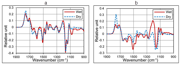
Fig. 2. Difference spectra of beech wood samples after 36 hours of UV treatment at 32°C (a) and at 53 °C (b) under wet and dry conditions
This deviation was strongly evidenced in the samples treated at 53 °C. It can be hypothesized that, under dry conditions, the beech wood samples underwent oxidation reactions attributable to free radicals generated by the UV light. When a certain amount of relative humidity was involved, these oxidation products reacted further with the water molecules, resulting in volatile low molecular compounds.
The liquid water is able to leach out the chemical compound-containing carbonyl groups, which absorb around 1767 cm-1. This effect is discussed later. Therefore, another explanation for the reduction of this band intensity is the presence of water molecules, which are able to remove carbonyl groups from the surface of the wood as well. At higher temperatures (53 °C), this phenomenon is more evident if compared to that observed at 32 °C. This can be attributed to an increase of molecule mobility with the temperature.
In the 1100 to 1300 cm-1 wavenumber region, both absorption increases and decreases were observed. The absorption increases were mainly generated by the irradiation in the wet condition. These were not real absorption rise values but can be associated with the observed roughness increase that causes an increment of the spectrum in this region. A detailed explanation of this phenomenon can be found in previous work (Tolvaj et al. 2011). This increment reduced the negative intensity of the band located at 1157 cm-1. The intensity of this negative band should increase with elapsed irradiation time, but after 16, 36, and 72 h of UV irradiation, the values of intensity were -0.18, -0.21, and -0.22. The roughness increase was determined by the optical method (Tolvaj et al. 2014). After 6, 16, 36, and 72 h of irradiation, these values were 1.31, 1.34, 1.38, and 1.4.
The spruce wood samples (Fig. 3) showed different behavior than did the beech wood. A similar decrease in the band at 1512 cm-1 was noticed, indicating the lignin degradation, but the absorption increase of the band at 1738 cm-1, assigned to carbonyl groups, differed considerably. The wet condition generated higher increases in the absorption of carbonyl groups than did the dry condition. The effect was more evident for the spruce wood samples treated at higher temperatures. This observation was the opposite of what had been observed for the beech wood. The 36 h UV treatment produced 1.6 times greater increase in absorption in the wet condition than in the dry condition for the samples treated at higher temperature. After 16 and 72 hours of treatment, this multiplication factor was 1.7 and 1.4, respectively. The decrease of this multiplication factor indicated the existence of the removal effect. The main difference between the IR spectra of beech and spruce is that there was no increase in the absorption peak at 1760 cm-1 for spruce.
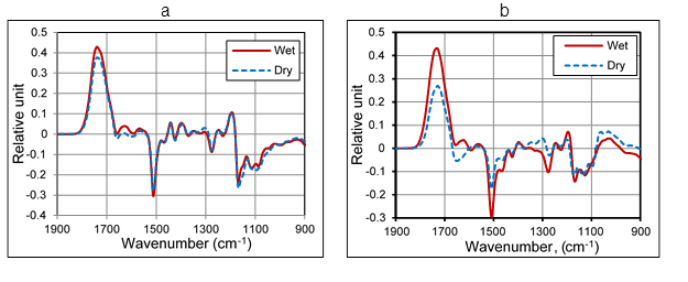
Fig. 3. Difference spectra of spruce wood samples after 36 hours of UV treatment at 32 °C (a) and at 53 °C (b) under wet and dry conditions
In Fig. 3b, a higher intensity decrease of the band at 1512 cm-1 was observed for the spruce samples treated at 53 °C in the dry rather than in the wet condition, while for the samples treated at 32 °C there was no difference in the decrease of the intensity of this band between the dry and wet conditions. This may indicate the combined contribution of water molecules and temperature for the increased effect of the photodegradation processes.
In order to clarify the leaching effect of water, some of the samples were inserted into distilled water after the UV treatment. After examining the photodegradation and leaching properties of the beech and spruce wood (Fig. 4), similar changes were found. The lignin content was not affected by the water leaching, as can be identified from the negative bands at 1596 and 1508 cm-1. Here, apparent differences are visible because of the baseline shift for beech. The photodegradation of extractives was indicated by the decrease of the band around 1650 cm-1. This conjugated carbonyl groups created by the photodegradation of extractives were leached out by the water (Fig.4/a).
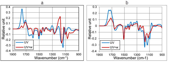
Fig. 4. Absorption change of beech (a) and spruce (b) after 1 day of UV treatment (UV) and 1 day of UV treatment followed by 1 day of water leaching (UV+w). (The initial spectrum was the reference in all cases.)
Two bands increased in intensity in the nonconjugated carbonyl region (1670 to 1820 cm-1) as a result of lignin degradation by the UV light. The stability of these two bands was not equal. The band at 1706 cm-1 was barely affected by the water leaching. However, the other band, at 1767 cm-1, was mostly removed, showing the same effect as the high humidity had produced.
The interpretation of the changes in the ether band region (1100 to 1200 cm-1) was difficult. The absorption intensities were calculated from the diffuse reflectance spectra using the K-M theory. But this theory does not work well in this region where the absorption of wood is strong. The increasing roughness tends to elevate the difference spectrum, confusing the real changes. The water leaching (and drying) produced an increase in roughness. The swelling lifted up the cut ends of the fibers. The drying did not bring them back, creating roughness increases at the micro level. This lifting effect is visible in Fig. 4.
CONCLUSIONS
- Spruce and beech samples were irradiated by UV light in humid and dry conditions. For comparison, the UV-irradiated samples were soaked in distilled water. The wet condition generated considerably greater discoloration than did the dry condition. The intensity of the color change was higher at the elevated temperature (53 °C) than at 32 °C.
- The results showed that the presence of water in the form of vapor increased the degradation of lignin. The nonconjugated carbonyl groups, absorbing at around 1760 cm-1, were not stable, and the number of these chemical groups decreased in the presence of vapor, in contrast to the dry condition. The increase in temperature amplified this degradation effect.
ACKNOWLEDGMENTS
This research, as a part of the Talentum Project for Science and Student Talent Fostering at WHU, TÁMOP-4. 2. 2. B-15/1/KONV-2015-0005, was sponsored by the EU/European Social Fund. The financial support is gratefully acknowledged.
REFERENCES CITED
Ayadi, N., Lejeune, F., Charrier, F., Charrier, B., and Merlin, A. (2003). “Color stability of heat-treated wood during artificial weathering,” Holz Roh Werkstoff 61, 221-226. DOI: 10.1007/800107-003-0389-2
Agresti, G., Bonifazi, G., Calienn, L., Capobianco, G, Lo Monaco, A., Pelosi, C, Picchio. R., and Serranti, S. (2013). “Surface investigation of photo-degraded wood by color monitoring, infrared spectroscopy, and hyperspectral imaging,” J. Spectrosc. 1(1), Article number 380536.
Bekhta, P., and Niemz, P. (2003). “Effect of high temperature on the change in color, dimensional stability and mechanical properties of spruce wood,” Holzforschung 57(5), 539-546.
Bonifazi, G., Calienno, L., Capobianco, G., Lo Monaco, A., Pelosi, C., Picchio, R., and Serranti, S. (2015). “Modelling color and chemical changes on normal and red heart beech wood by reflectance spectrophotometry, Fourier Transform infrared spectroscopy and hyperspectral imaging,” Polym. Degrad. Stab. 113, 10-21.
Butler, D. A., Brunner, C. C., and Funck, J. W. (2001). “Wood-surface feature classification using extended-color information,” Holz Roh Werkstoff 59(6), 475-482.
Calienno, L., Lo Monaco, A., Pelosi, C., and Picchio, R. (2014). “Clour and chemical changes on photodegraded beech wood with or without red heartwood,” Wood Sci. Technol. 48(6), 1167- 1180.
Chang, H. T., and Chang, S. T. (2001). “Correlation between softwood discoloration induced by accelerated lightfastness testing and indoor exposure,” Polym. Degrad. Stab. 72, 361-365. DOI: 10.1016/S0141-3910(01)00039-8
Chang, T. C., Chang, H. T., Wu, C. L., and Chang, S. T. (2010). “Influences of extractives on the photodegradation of wood,” Polym. Degrad. Stab. 95, 516-521. DOI: 10.1016/j.polymdegradstab.2009.12.024
Colom, X., Carrillo, F., Nogués, F., and Garriga, P. (2003). “Structural analysis of photodegraded wood by means of FTIR spectroscopy,” Polym. Degrad. Stab. 80, 543-549. DOI: 10.1016/S0141-3910(03)00051-X
Esteves, B., Marques, A. V., Domingos, I., and Pereira, H. (2008). “Heat-induced colour changes of pine (Pinus pinaster) and eucalypt (Eucalyptus globulus) wood,” Wood Sci. Technol. 42(5), 369-384.
Fan, Y., Gao, J., and Chen, Y. (2010). “Colour responses of black locust (Robinia pseudoacacia L.) to solvent extraction and heat treatment,” Wood Sci. Technol. 44, 667-678. DOI: 10.1007/s00226-009-0289-7
George, B., Suttie, E., Merlin, A., and Deglise, X. (2005). “Photodegradation and photostabilisation of wood – the state of the art,” Poly. Degrad. Stab. 88(2), 268-274.
Gou, M., and Guan, X. (2010). “Effect of UV radiation on surface color and chemical structure of wood,” Adv. Mat. Res. 113-116, 1624-1628. DOI: 10.4028/www.scientific.net/AMR.113-116.1624
Hon, D. N. S., and Minemura, N. (2001). “Color and discoloration,” in: Wood and Cellulosic Chemistry, 2nd Ed., rev. and expanded, Hon, D. N. S., and Shiraishi, N. (eds.), pp. 385-442, Marcel Dekker, New York
Matsuo, M., Yokoyama, M., Umemura, K., Gril, J., Yano, K., and Kawai, S. (2010). “Color changes in wood during heating: Kinetic analysis by applying a time-temperature superposition method,” Appl. Phys. A 99(1), 47-52.
Miklečić, J., Jirous-Rajković, V., Antonović, A., and Španić, N. (2011). “Discoloration of thermally modified wood during simulated indoor sunlight exposure,” BioResources 6, 434-446.
Müller, U., Rätzsch, M., Schwanninger, M., Steiner, M.,and Zöbl, H. (2003). “Yellowing and IR changes of spruce wood as result of UV-irradiation,” J. Photochem. Photobiol. B: Biol. 69(2), 97-105.
Nzokou, P., and Kamdem, P. D. (2006). “Influence of wood extractives on the photo-discoloration of wood surfaces exposed to artificial weathering.” Ind. Appl. 31, 425-434.
Oltean, L., Teischinger, A., and Hansmann, C. (2008). “Wood surface discolouration due to simulated indoor sunlight exposure,” Holz Roh Werkstoff 66, 51-56. DOI: 10.1007/s00107-007-0201-9
Oltean, L., Hansmann, C., Nemeth, R., and Teischinger, A. (2010). “Wood surface discolouration of three Hungarian hardwood species due to simulated indoor sunlight exposure,” Wood. Res. Slov. 55, 49-58.
Pandey, K. K. (2005a). “Study of the effect of photo-irradiation on the surface chemistry of wood,” Polym. Degrad. Stab. 90, 9-20. DOI: 10.1016/j.polymdegradstab.2005.02.009
Pandey, K. K. (2005b). “A note on the influence of extractives on the photo-discoloration and photo-degradation of wood,” Polym. Degrad. Stab. 87, 375-379. DOI: 10.1016/j.polymdegradstab.2004.09.007
Persze, L., and Tolvaj, L. (2012). “Photodegradation of wood at elevated temperature: Colour change,” J. Photochem. Photobiol. B: Biol.108, 44-47. DOI: 10.1016/j.jphotobiol.2011.12.008.
Popescu, C. M., Popescu, M. C., and Vasile, C. (2011). “Structural analysis of photodegraded lime wood by means of FT-IR and 2D IR correlation spectroscopy,” Int. J. Biol. Macromol. 48, 667-675. DOI: 10.1016/j.ijbiomac.2011.02.009.
Rosu, D., Teaca, C. A., Bodirlau, R., and Rosu, L. (2010). “FTIR and color change of the modified wood as a result of artificial light irradiation,” J. Photochem. Photobiol. B: Biol. 99, 144-149. DOI: 10.1016/j.jphotobiol.2010.03.010
Schnabel, T., Zimmer, B., and Petutschnigg, A. J. (2009). “On the modeling of colour changes of wood surfaces,” Holz Roh Werkstoff 67(2), 141-149.
Sharratt, V., Hill, C. A. S., and Kint, D. P. R. (2009). “A study of early colour change due to simulated accelerated sunlight exposure in Scots pine (Pinus sylvestris),” Poly.Degrad. Stab. 94(9), 1589-1594.524.
Srinivas, K., and Pandey, K. K. (2012). “Photodegradation of thermally modified wood,” J. Photochem. Photobiol. B: Biol. 117, 140-145. DOI: 10.1016/j.jphotobiol.2012.09.013
Teacǎ, C. A., Roşu, D., Bodîrlǎu, R., and Roşu, L. (2013). “Structural changes in wood under artificial UV light irradiation determined by FTIR spectroscopy and color measurements-a brief review,” Bioresources 8(1), 1478-1507.
Tolvaj, L., and Faix, O. (1995). “Artificial ageing of wood monitored by DRIFT spectroscopy and CIE L*a*b* color measurements. I. Effect of UV light,” Holzforschung 49, 397-404.
Tolvaj, L., Mitsui, K., Varga, D. (2011). “Validity limits of Kubelka–Munk theory for DRIFT spectra of photodegraded solid wood,” Wood Sci. Technol. 45, 135-146. DOI: 10.1007/s00226-010-0314-x
Tolvaj, L., Molnar, Zs., Nemeth, R. (2013). “Photodegradation of wood at elevated temperature: Infrared spectroscopic study,” J. Photochem. Photobiol. B: Biol. 121, 32-36. DOI: 10.1016/j.jphotobiol.2013.02.007
Tolvaj, L., Molnar, Zs., and Magoss, E. (2014). “Measurement of photodegradation-caused roughness of wood using a new optical method,” J. Photochem. Photobiol. B: Biol. 134, 23-26. DOI: 10.1016/j.jphotobiol.2014.03.020
Tolvaj, L., Tsuchikawa, S., Inagaki, T., and Varga, D. (2015). “Temperature dependence of photodegradation of wood monitored by colour measurement,” Wood Sci. Technol. 49, 1225-1237. DOI: 10.1007/s00226-015-0749-1
Xie, Y., Krause, A., Mai, C., Militz, H., Richter, K., Urban, K., and Evans, P. D. (2005). “Weathering of wood modified with the N-mrthylol compound 1,3-dimethylol-4,5-dihydroxyethyleneurea,” Polym. Degrad. Stab. 89, 189-199. DOI: 10.1016/j.polymdegradstab.2004.08.017
Zahri, S., Belloncle, C., Charrier, F., Pardon, P., Quideau, S., and Charrier, B. (2007). “UV light impact on ellagitannins and wood surface colour of Europena oak (Quercus petraea and Quercus robur),” Appl. Surf. Sci. 253, 4985-4989.
Zivkovic, V., Arnold, M., Radmanovic, K., Richter, K., and Turkulin, H. (2013). “Spectral sensitivity in the photodegradation of fir wood (Abies alba Mill.) surfaces: Colour changes in natural weathering,” Wood Sci. Technol. 48(2), 239-252. DOI: 10.1007/s00226-013-0601-4
Article submitted: July 29, 2015; Peer review completed: October 24, 2015; Revised version received and accepted: October 30, 2015; Published: November 16, 2015.
DOI: 10.15376/biores.11.1.296-305