
Abstract
Melanins are complex molecules found in many organisms including bacteria, fungi, plants, and animals that have protective functions against stress. In fungi there are several biochemical routes for their synthesis, and their diversity and structural complexity have made their analysis and characterization difficult. However, the possible specific functions of melanins in organisms have been determined. Based on their physicochemical properties, their potential in the application in various areas of interest and benefit of the human being have been visualized, such as in health and medicine, in bioremediation, and in the food industry. In this review, the type of melanins produced by fungi are discussed, as well as their main biological functions, the main biochemical routes involved in their synthesis, and potential applications in various areas. An important research area is visualized to find the best melanin-producing fungi as well as the conditions in which their production is maximized, in order to continue investigating the relationship of the structure of melanin with its biological functions as well as the determination of its physicochemical properties to establish its applications.
Download PDF
Full Article
Fungal Melanins and their Potential Applications: A Review
Sac N. Salgado-Castillo,a Hugo A. López-Peña,b Rubén Díaz,c Kathia Peña-Solis,c Edith Ponce-Alquicira,a Jorge Soriano-Santos,a,* and Gerardo Díaz-Godínez c,*
Melanins are complex molecules found in many organisms including bacteria, fungi, plants, and animals that have protective functions against stress. In fungi there are several biochemical routes for their synthesis, and their diversity and structural complexity have made their analysis and characterization difficult. However, the possible specific functions of melanins in organisms have been determined. Based on their physicochemical properties, their potential in the application in various areas of interest and benefit of the human being have been visualized, such as in health and medicine, in bioremediation, and in the food industry. In this review, the type of melanins produced by fungi are discussed, as well as their main biological functions, the main biochemical routes involved in their synthesis, and potential applications in various areas. An important research area is visualized to find the best melanin-producing fungi as well as the conditions in which their production is maximized, in order to continue investigating the relationship of the structure of melanin with its biological functions as well as the determination of its physicochemical properties to establish its applications.
DOI: 10.15376/biores.18.4.Castillo
Keywords: Biological activity; Biosynthesis pathways; Fungi; Melanin
Contact information: a: Department of Biotechnology, Metropolitan Autonomous University-Iztapalapa, Mexico City, 09340, Mexico; b: Chemistry Department, Virginia Commonwealth University, Richmond 23284, USA; c: Research Center for Biological Sciences, Autonomous University of Tlaxcala, Tlaxcala, 90000, Mexico; *Corresponding author: gerardo.diaz@uatx.mx; jss@xanum.uam.mx
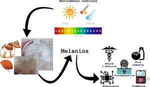
GRAPHICAL ABSTRACT
INTRODUCTION
The term melanin originates from the Greek word “melanos”, which means black (Gessler et al. 2014). Melanin is the broad term used to refer to a group of dark-colored natural pigments. Melanin is produced by many organisms, including bacteria, fungi, plants, and animals (Ghattavi et al. 2022). There is fossil evidence of the presence of melanin in various organisms (Zhang et al. 2010; Glass et al. 2012). Melanin is a pigment that, from a chemical point of view, is a heterogeneous, hydrophobic polymer of high molecular weight, indigestible, negatively charged, and derived from the oxidation of phenolic or indole type of compounds; these structures lead to intermediate phenols including quinones that subsequently polymerize (Solano 2014; Suwannarach et al. 2019). In general, four types of melanin are considered: eumelanin, pheomelanin, neuromelanin, and allomelanin, which are based mainly on the chemical precursors for their biosynthesis. However, although they have been studied for a long time, their chemical composition has not been precisely determined, so not all of their functions and properties have been characterized (Riley 1997; Cao et al. 2021).
The subgroup of melanin with coloration from black to brown is called eumelanin, which is the most common in animals, including humans, although it is also produced by fungi and bacteria. This melanin is one of the most studied. It is produced from the oxidative polymerization of tyrosine derivatives such as L-3,4-dihydroxyphenylalanine (L-Dopa) (Langfelder et al. 2003; Solano 2014; Tran-Ly et al. 2020a). Pheomelanin is a melanin with red or yellow coloration, of animal origin, mainly in mammals and birds. This melanin is found in red hair, freckles, or feathers. It is synthesized from the same precursor as eumelanin but incorporates a cysteine to L-DOPA (5-S-cysteinyl-Dopa). Neuromelanin is produced within human neurons through the oxidation of dopamine, norepinephrine, and catecholamines (Hirsch et al. 1988; Zecca et al. 2002). Allomelanin (other melanins) is a pigment found in plants and fungi that does not contain nitrogen. Given the research on animal melanin, it was considered that this type of compound should contain nitrogen in its molecule; therefore, for a long time, this plant pigment was not considered as melanin. However, research on black pigments obtained from animals, plants, and microorganisms, shows common physicochemical properties, except for the absence of nitrogen in those of plant origin. Thus, currently the term melanin does not include the requirement of nitrogen as part of the molecule (Solano 2014; Glagoleva et al. 2020). The allomelanin group is the most heterogeneous. There are many precursors for its synthesis. Pyomelanin and DHN-melanin are melanins without nitrogen and are found in bacteria and fungi. The latter can be formed from γ-glutaminyl-3,4-dihydroxybenzene, catechol, and 1,8-dihydroxynaphthalene. In contrast, the catechol-melanin found in plants has catechol, and the caffeic, chlorogenic, protocatechuic, and gallic acids as precursors. It is worth mentioning that many microorganisms could produce different types of melanin, including eumelanin, using a synthesis pathway similar to that observed in mammals (Solano 2014; Glagoleva et al. 2020; Tran-Ly et al. 2020a).
Among the most important functions of melanin is the protection of the organism from ultraviolet radiation (UV) because it has the property of absorbing UV light, thus minimizing cell damage (Eisenman and Casadevall 2012; Gessler et al. 2014; Braga et al. 2015). In humans, there are melanocytes in the basal layer of the epidermis. These are the cells where melanin is produced. Such melanin is responsible for skin pigmentation; thus there is a greater amount of melanin in people with darker skin. In addition, its content can be increased by exposure to UV light, for example as a consequence of tanning the skin (Natarajan et al. 2014). Various functions of melanin have been reported in different organisms, for example, in insects, melanin has several functions, including pigmentation of its exoskeleton, hardening of the cuticle; it is involved in wound healing and promotes the immune response (Sugumaran and Barek 2016). In organisms, such as octopuses, cuttlefish, cephalopods, and squids, melanin serves as protection against predators (Derby 2014); in mollusks, with the exception of cephalopods, melanin is part of the pigments they use to color their shells (Williams 2016). In birds, melanin primarily gives plumage coloration (Galvan and Solano 2016). In amphibians, pigmentation by eumelanin occurs, in some cases, in the brain for protection against UV radiation, changes in osmolarity and temperature, in the case of Xenopus laevis melanin by protecting against UV light, prevents damage at the level of nervous system and possible mutagenic alterations (Díaz and Pedemonte 2013). In bacteria, such as Vibrio cholerae, melanin has been associated with protective properties at high temperatures and high solute concentrations (Ivins and Holmes 1981; Kotob et al. 1995). In fungi, melanin has been related to longevity, pathogenicity, and survival (Sussman et al. 1963; Wheeler and Bell 1988), among which the most important functions is protection against adverse environmental conditions, even calling melanin “fungal armor” (Pombeiro-Sponchiado et al. 2017). It protects fungi against changes in osmolarity (Mattos et al. 2020; Yaakoub et al. 2021), high temperatures (Gessler et al. 2014), bacterial attack (Nosanchuk and Casadevall 2003), as well as provides radioprotection and photoprotection (Pombeiro-Sponchiado et al. 2017). Melanin has effects on virulence in pathogenic fungi, protecting them from mechanism’s host immune defense (Chongkae et al. 2021; Mattoon et al. 2021). It has also been observed that melanin may have a resistance effect to antifungals (e.g., amphotericin B, flucytosine, miconazole, ketoconazole, fluconazole, and itraconazole) (Ruíz-Diez and Martínez-Suárez 2003; Waller et al. 2021). Melanin can have an immunomodulatory effect on a host organism, causing an antibody production response (Wong et al. 2018; Mora-Montes 2020). In addition, it presents antimicrobial, antiviral, cytotoxic, anti-inflammatory, and immunomodulatory activity (Pombeiro-Sponchiado et al. 2017). The melanin produced by Octopus mimus showed growth inhibition of Staphylococcus aureus and Escherichia coli (Vega-Petkovic 2013). Melanin also can act as an effective antioxidant as it provides protection against oxidative stress, free radicals, and oxidizing agents (Lin and Chen 2005; Meredith and Sarna 2006; Aseervatham et al. 2013; Maher et al. 2020).
In this review, the types of melanins produced by fungi are discussed, as well as their known biological functions and the main biochemical pathways involved in their synthesis. Also considered are studies in which, based on melanins’ physicochemical properties, their potential applications have been suggested in various areas such as bioremediation, medicine, health, food, and even electronics.
FUNGAL MELANINS
Melanins are not essential for the growth and development of fungi; however, they are secondary metabolites that are produced as survival mechanisms under extreme conditions of pH, temperature, salinity, and the presence of radioactivity. They also can confer virulence in the pathogenicity (Belozerskaya et al. 2017). It is worth mentioning that there is still much to be investigated regarding the structure, synthesis, and function of melanins in fungi, to take advantage of their properties in various biotechnological applications, including their ability to counteract fungal infections (Eisenman et al. 2020). Microscopic studies show that in fungi, melanin occurs in the form of granules located in the cell wall, although its distribution (in the outer or inner layer of the cell wall) may vary between species (Nosanchuk and Casadevall 2003; Morris-Jones et al. 2005; Walker et al. 2010). The cell walls of fungi are made up of glucans and chitin in the form of linked fibers, where the material primarily contains proteins and mannans (Eisenman and Casadevall 2012). The outer layer contains high levels of different types of mannoproteins, while the inner layer is composed mostly of polysaccharides (beta-glucans and chitin) and small amounts of protein. It has been suggested that fungal melanin can be synthesized in internal vesicles similar to melanosomes in animal tissues, and from there they are transported to the cell wall (Eisenman and Casadevall 2012; Belozerskaya et al. 2017). In the pathogenic fungus Fonsecaea pedrosoi, there are vesicles that have a fibrillar matrix, like that present in amphibian melanosomes. The vesicles act as a site for melanin accumulation, which is then transported to the cell wall and deposited in concentric layers (Franzen et al. 2008). Various studies have shown that melanins have specific functions depending on the species of fungus. It has been observed that cell walls with melanin are less porous than non-melanized ones, so the incorporation of melanin is a fungal strategy to regulate the pore sizes of their cell walls, participating in their mechanical strength, resulting in a defense system against harmful environments, as well as an interaction factor with the host and parasites that could attack the fungus (Nosanchuk and Casadevall 2003; Nosanchuk et al. 2015). It was determined that melanin promotes resistance to osmotic stress and desiccation in Cenococcum geophilum (Eisenman et al. 2020).
It has been reported that melanins in fungi also participate in increasing the virulence of parasitic fungi, as in the case of Sporothrix schenckii, Paracoccidioides brasiliensis, and Exophiala (Wangiella) dermatitidis; it contributes to fungal pathogenesis by altering the host defense response mechanisms, thus decreasing phagocytosis as in the case of Cryptococcus neoformans and Penicillium marneffei (Eisenman et al. 2020). They also protect fungi against fungicidal peptides and antifungal drugs and have antioxidant activity (Schnitzler et al. 1999; Romero-Martinez et al. 2000; Nosanchuk et al. 2015). Melanins can also absorb UV radiation, avoiding its harmful effects and overheating, so that they are excellent photoprotectors in fungi (Belozerskaya et al. 2017). Melanin also protects fungi against radioactive contamination. It has been observed that there is a high incidence of melanized fungi including species, such as Cladosporium spp., Alternaria alternata, Aureobasidium pullulans, and Hormoconis resinae, growing in the area where the Chernobyl nuclear reactor was damaged, where there is constant exposure to high radiation levels (Gessler et al. 2014).
There is scientific evidence that melanized fungi of the Cladosporium and Alternaria genera are more abundant in industrial areas and on highways because they are more resistant to contamination by heavy metals and unsaturated hydrocarbons (Dadachova et al. 2008; Dighton et al. 2008; Gessler et al. 2014). In one study, it was observed that in a melanized cell (in the presence of L-DOPA) of P. marneffei, the drugs amphotericin B, clotrimazole, ketoconazole, itraconazole, and fluconazole were less effective compared to non-melanized cells (Kaewmalakul et al. 2014). A similar effect was observed in Scedosporium prolificans, where melanin showed a protective effect and resistance to antifungals such as amphotericin B, flucytosine, miconazole, ketoconazole, fluconazole, and itraconazole (Ruíz-Diez and Martínez-Suárez 2003; Urán and Cano 2008). It was also observed that melanin is responsible for resistance to amphotericin B in Blastomyces dermatitidis, in C. neoformans, and Histoplasma capsulatum for resistance to amphotericin B, fluconazole, and caspofungin, and for P. brasiliensis it showed resistance to the action of amphotericin B, fluconazole, ketoconazole, and itraconazole (Urán and Cano 2008).
Most ascomycetes and deuteromycetes synthesize melanin from 1,8-dihydroxy-naphthalene (DHN). Some basidiomycetes synthesize it from L-3,4 dihydroxyphenyl-alanine (L-DOPA) or glutaminyl-3,4-dihydroxybenzene (GDHB) (Henson et al. 1999; Selvakumar et al. 2008). Many fungi never produce any form of melanin under normal growth conditions, but in the presence of L-DOPA by spontaneous oxidation they will produce DOPA-melanin under slightly alkaline conditions without enzymatic intervention. However, in fungi that are not melanin producers, with the participation of enzymes, such as laccase, some peroxidases and/or catalases, can form black polymers from L-DOPA (Butler and Day 1998). Research on the black yeast Phaeococcomyces sp. showed that it produces melanin of DHN origin and that mutants of this yeast darkened when they were put in contact with a solution of L-DOPA (Butler et al. 1989). The yeast C. neoformans causes potentially fatal meningoencephalitis in 5% to 10% of AIDS patients. It can form DOPA-melanin from L-DOPA, a brain neurotransmitter (Wang and Casadevall 1994). In this same yeast, laccase has been observed to catalyze the formation of melanin by oxidizing L-DOPA, initiating a series of reactions that lead to pigment polymerization in the yeast cell wall (Butler et al. 1989).
PATHWAYS OF MELANIN SYNTHESIS IN FUNGI
As previously mentioned, melanins are considered secondary metabolites that are basically a protection mechanism that fungi have against various adverse environmental conditions. Therefore, intrinsic and extrinsic factors that cause stress to fungi favor their production. UV radiation is a factor that increases melanin production, so it has also been observed that radioactivity also promotes its production. The growth temperature of fungi influences their melanin production capacity. In many cases, the optimal growth temperature is not the same as the temperature where pigment production is maximized. The pH is another important factor in melanin production, influencing the amount and type of pigment, since the same fungus can produce different pigments by changing the pH of the culture medium. On the other hand, it has been observed in some fungi that light inhibits the synthesis of melanins that are produced when fungi grow in the dark; aeration as well as the type and quantity of carbon and nitrogen sources are determinants in the growth and production of melanins (Pombeiro-Sponchiado et al. 2017).
Various melanin biosynthesis pathways have been identified in fungi from phenolic and indole precursors. The acetate-malonate pathway is one of the most common and widely studied, where acetyl and malonyl coenzyme A are the building blocks for the synthesis of polyketides through a series of Claisen condensations and successive decarboxylation (Ibrahim and Mohamed 2016). This polyketide synthesis, assisted by polyketide synthase enzymes, is involved in the DHN-melanin synthesis pathway (Butler and Day 1998; Gessler et al. 2014; Solano 2014; Toledo et al. 2017) and also in the synthesis of the recently discovered deoxybostrycoidin-melanin (Frandsen et al. 2016) (Fig. 1). There are some variations between the polyketides found as precursors in the synthesis of melanins in different species of fungi, but in general they can be categorized as penta- and hepta-ketides (Tsai et al. 2001). In DHN-melanin biosynthesis, the polyketide precursor is converted to 1,3,6,8-tetrahydroxynaphthalene (THN). Subsequently, due to the action of hydroxynaphthalene reductase, scytalone is formed, which is dehydrated to form 1,3,8-trihydroxynaphthalene that is then reduced to vermelone and through an additional dehydration step, 1,8-dihydroxynaphthalene (DHN) is generated. The DHN is polymerized to form DHN-melanin with the participation of enzymes, such as laccase, or some other type of polyphenol oxidase (Fig. 1). In general, the structure of the DHN-melanin polymer is not known in detail, but the DHN-melanin in the mycelium of the fungus Mycosphaerella fijiensis consists of approximately 50 DHN units (Beltrán-García et al. 2014). It is possible that DHN-melanin synthesis starts from glucose when acetyl-CoA is formed, but if glucose enters the pentose pathway, L-DOPA can be formed to continue along the tyrosine pathway where eumelanins and pheomelanins are obtained (Urán and Cano 2008). Frandsen et al. (2016) identified that the perithecial black pigmentation in Fusarium graminearum is due to the accumulation of melanin derived from 5-deoxybostrycoidin. Based on the various chemical structures and enzymes identified and involved in melanin synthesis, the authors have made a proposal for the deoxybostrycoidin-melanin synthesis pathway. Said route also starts from acetyl-CoA and malonyl-CoA, which is shown in a simplified way in Fig. 1. It is suggested that this type of melanin may be relevant for other Fusarium species with dark perithecium.

Fig. 1. Biosynthesis of deoxybostrycoidin-melanin and DHN-melanin in fungi (Adapted from Frandsen et al. 2016; Almeida-Paes et al. 2017)
There is evidence that fungi produce melanin from glutaminyl-4-hydroxybenzene (GHB) synthesized from derivatives of the shikimate pathway (Jadot et al. 1960; Jolivet et al. 1998) (Fig. 2).

Fig. 2. Biosynthesis of GHB-melanin and PAP-melanin in fungi (Adapted from Weijn et al. 2013; Lino and Manini 2022)
From shikimate, through a series of transformations, chorismate is formed. This is converted into p-aminobenzoate and later into p-aminophenol (PAP) to finally give rise to GHB. The GHB then is oxidized to glutaminyl-3,4-dihydroxybenzene (GDHB) with the participation of a tyrosinase.
Some results indicate that GHB, tyrosine, and the products derived from their hydrolysis and/or oxidation are the most abundant phenolic compounds in Agaricus bisporus (Jolivet et al. 1998). The GHB and some of its oxidation products are believed to be the building blocks that give rise to so-called GHB-melanin (Butler and Day 1998). A. bisporus is also capable of synthesizing PAP-melanin (Fig. 2). The synthesis begins from the conversion of PAP into ρ-aminocatechol, after a series of polymerization steps this substrate is transformed into PAP-melanin (Weijn et al. 2013).
It is possible to find more melanins coming from metabolites of the shikimic acid pathway (Fig. 3). Chorismate is converted to prephenate by the action of chorismate mutase. The prephenate is transformed into p-hydroxyphenyl pyruvate with the help of prephenate dehydrogenase, and then tyrosine is obtained. Tyrosine can be oxidized to a lesser extent, to give rise to L-DOPA, or to a greater extent, to generate dopaquinone (DAQ). The oxidation of L-DOPA to DAQ is also possible (Langfelder et al. 2003). This series of oxidations beginning with tyrosine is catalyzed by some type of polyphenol oxidase. The formation of DAQ is followed by a cyclization that gives rise to leucodopachrome. This compound is oxidized to dopachrome in the presence of DAQ, and finally 5,6-dihydroxyindole-2-carboxylic acid (DHICA) is obtained. However, if there is decarboxylation at position 2 of the indole, it will form 5,6-dihydroxyindole (DHI). It is believed that this decarboxylation can take place at some point in the pathway after DAQ generation, and for that reason in Fig. 3 the carboxylate group is shown in parentheses in that species.
Both DHICA and DHI can polymerize to form DOPA-melanin (black and brown eumelanins) (Gessler et al. 2014; Almeida-Paes et al. 2017; Toledo et al. 2017). It has been shown that the variation in the concentration of copper in the culture medium of a fungus can cause changes in its pigmentation (Eisenman and Casadevall 2012). This is because copper is extremely important for the synthesis of DHN-melanin, DOPA-melanin, and other melanins. The importance lies in the fact that copper functions as a cofactor of the oxidase enzymes that participate in the biosynthesis of melanin.
In fungi, the synthesis of pheomelanins from tyrosine is also possible, more specifically from its oxidation product, dopaquinone (Fig. 3). Dopaquinone reacts with cysteine or glutathione to form cysteinyl-DOPA. Subsequently, the cyclization of the product ends in a benzothiazine derivative, whose polymerization leads to the formation of yellow, brown, or red pigments known as pheomelanins (Gessler et al. 2014; Solano 2014). Fungi have a large enzymatic machinery capable of forming melanins through other pathways from tyrosine (Fig. 3). Through a reaction catalyzed by tyrosine transaminase, p-hydroxyphenylpyruvate is formed, which is converted by dioxygenase into homogentisate, which is spontaneously oxidized to benzoquinone acetate. This last compound is polymerized, forming soluble brown pigments called pyomelanins (Gessler et al. 2014; Solano 2014). Geib et al. (2016) reported that Aspergillus terreus produces asp-melanin from the polymerization of aspulvinone E, revealing another example of melanin generated from tyrosine (Fig. 3).
Finally, there is a little-studied pathway that starts from the catechol, a substrate that has been identified in the tissue of the fungi and derives in a catechol-melanin. Until now, the metabolic pathway for the synthesis of this melanin is not completely known; however, Fig. 4 shows a proposal for the synthesis route.

Fig. 3. Biosynthesis of pyomelanin, asp-melanin, DOPA-melanin, and pheomelanin in fungi. (Adapted from Weijn et al. 2013; Geib et al. 2016; Almeida-Paes et al. 2017; Eisenman et al. 2020; Lino and Manini 2022). NRPS: non-ribosomal peptide synthetase
It should be mentioned that there are some compounds that can inhibit the synthesis of DOPA-melanin, such is the case of tropolone, kojic acid, 2-mercaptobenzimidazole, and diethyldithiocarbamate. Regarding the inhibition of DHN-melanin, there are tricyclazole, pyroquilone, phthalide, and clobenthiazone (Selvakumar et al. 2008).

Fig. 4. Biosynthesis of catechol-melanin in fungi (Adapted from Solano 2014; Ito et al. Wakamatsu 2020; Cao et al. 2021)
Despite the complications in the chemical identification of melanins, there are efforts to characterize them as far as possible based on their chemical behavior. The chemical criteria that are generally used to define a fungal melanin are the following: dark coloration, insolubility in cold or hot water and in organic solvents, resistance to degradation by hot or cold concentrated acids, bleaching by oxidizing agents (e.g., hydrogen peroxide), ability to directly reduce ammoniacal silver nitrate solutions, and solubilization and degradation in hot alkaline solutions (Meredith and Sarna 2006).
These recalcitrant properties of melanins have been widely used to purify them. The treatment of samples with hot and acid solvents has been used to degrade the rest of the biological structures present in the sample, assuming that the residue corresponds to melanin (Gómez and Nosanchuk 2003).
POTENTIAL APPLICATIONS OF FUNGAL MELANINS
Based on the knowledge of the physicochemical properties and biological functions of melanins, their potential biotechnological use has been determined. Research has suggested the main areas where they can impact, including medicine, in environmental remediation processes, and in various industrial applications. Because melanins have antioxidant, immunomodulatory, photoprotective, radioprotective, anti-inflammatory, hypoglycemic, liver protective, and gastrointestinal benefits, among others, they have potential applications in the health area, in the pharmaceutical industry, and in biomedicine (ElObeid et al. 2017). Cosmetic companies have used melanins in the formulation of creams and sunscreens because melanin has high UV light absorption and antioxidant activity. The sun protection performance, antioxidant activity, and cytotoxicity of the melanin produced by the ascomycete Amorphotheca resinae was recently determined. Subsequently, melanin (5%) was part of the ingredients of a cream, showing an in vitro sun protection factor value of 2.5. Moreover, it showed a critical wavelength of about 388 nm and a UVA/UVB ratio of more than 0.81, which is satisfactory to cover the requirement of broad-spectrum sun protection. The antioxidant activity of melanin was similar to that presented by ascorbic acid and higher than that of reduced glutathione. It was not cytotoxic to human keratinocyte cell lines up to 72 h of exposure (up to a concentration of 4 mg melanin/mL) (Oh et al. 2021a). Among the properties of melanins is to provide radioprotection to fungi, so there is the possibility that they can function as radioprotectors of the human intestine in people who receive radiation therapy. Pacelli et al. (2017) compared the effect of highly ionizing deuterons (deuterium nuclei) and X-rays (ionizing to a lesser extent than deuterons) on melanized and non-melanized varieties of C. neoformans and Cryomyces antarcticus, microscopic fungi capable of forming DOPA- and DHN-melanin, respectively. Melanin conferred protection against high doses of deuterons (1.5 kGy) to both fungi, although this effect was more significant for C. neoformans. As for X-rays, at a dose of 0.3 kGy, melanin exerted a protective effect for C. antarcticus but not for C. neoformans. Revskaya et al. (2012) used two groups of mice; one group was fed with the edible black mushroom Auricularia auricula-judae (melanized mushroom) and the other with the white mushroom Boletus edulis (without melanin). The mice were irradiated with cesium 137 at a dose of 9 Gy and a radiation flux of 2.5 Gy/min, for 45 days. After 14 days of irradiation, 80% of the mice fed with white fungus had died, while 60% of those fed with black fungus survived until the end of the experiment. The number of leukocytes found in the blood of the surviving mice fed with black fungus did not present a significant difference with that of non-irradiated mice. A lower concentration of platelets was quantified, and there was no damage in the stomach, or large and small intestines. In another study, using mice, the radioprotective effect of extracellular melanin from the fungus Gliocephalotrichum simplex was evaluated. The mice supplied with melanin (50 mg/kg of body weight) were irradiated with a dose of 7 Gy, managing to increase their survival. Melanin inhibited radiation-induced hematopoietic damage, also reduced oxidative stress in liver tissue, and eliminated immune imbalance by reducing the production of proinflammatory cytokines (Kunwar et al. 2012). Based on the protective property of melanin against radiation, studies have been conducted for its use in protecting astronauts from space radiation (Cordero 2017).
In another study, it was reported that the halotolerant black marine yeast Hortaea werneckii AS1 isolated from the Mediterranean salt lakes in Alexandria, produced melanin with antioxidant activity assessed by the DPPH method with an IC50 of 61.38 μg/mL. It did not show cytotoxicity towards the human skin fibroblast cell line (IC50 > 0.1 mg/mL). It is worth mentioning that it presented antimicrobial activity against Aeromonas sp. and Aspergillus niger (Elsayis et al. 2022). In another investigation, melanin was extracted from the residues of the Auricularia auricula culture, and its antioxidant activity (by the DPPH and ABTS methods) and some physicochemical properties were determined. This melanin presented antioxidant activity and also significantly inhibited cell death caused by H2O2, restoring cell viability to 98.09% when the melanin concentration was 1.6 mg/mL. It also showed the ability to improve the morphology of cells under stress oxidative induced (Liu et al. 2019). Recently, Hou et al. (2021) evaluated in vitro and in vivo the effect of A. auricula melanin on liver damage caused by alcohol. Healthy human liver cells were pretreated with ethanol and then treated with melanin. It was observed that melanin significantly improved cell viability and morphology and reduced the amount of reactive oxygen species and GSH/GSSG. Contrastingly, mice were given ethanol to induce acute alcoholic liver damage, and then melanin was given. It was observed that melanin in mice with liver damage caused a decrease in triglyceride and malondialdehyde levels and in the activities of alanine aminotransferase and aspartate aminotransferase, simultaneously the levels of cytosolic alcohol dehydrogenase, superoxide dismutase, and catalase were increased. The authors suggest that the therapeutic effect of melanin may be related to the inhibition of Cytochrome P450 2E1 (CYP2E1) expression and the activation of nuclear factor-E2 related factor 2 (Nrf2) and its downstream antioxidase.
Another potential application of melanin is as a nanocarrier for drugs, protecting them and promoting their release. In a study, melanin nanoparticles impregnated with the antibiotic metronidazole were created. The release rate of the antibiotic was determined at acidic and physiological pH, noting that the process is strongly dependent on pH. This, coupled with the biocompatibility and lack of cytotoxicity of melanin, could be a targeted drug nanocarrier system in the colon and intestine (Araújo et al. 2014). Lee and Hyun (2014) suggested that the water-soluble melanin from Inonotus obliquus could be an antidiabetic agent, as it has antihyperglycemic and beneficial effects on lipid metabolism. This was based on an investigation they conducted using obese mice supplied with melanin and after treatment observed improvement in insulin sensitivity and reduction in adiposity.
There are reports on the potential use of melanin to treat disorders of the immune system including AIDS, against different types of malignant cancerous tumors, in diseases of blood origin and disorders due to alterations in cellular homeostasis, also for complex mental disorders (e.g., schizophrenia, epilepsy, and Alzheimer’s disease) and those that affect the central nervous system such as Parkinson’s (Pombeiro-Sponchiado et al. 2017). Melanins also have a potential application in tissue regeneration, including neural and cardiac tissues. In one study, the use of melanin thin films enhanced Schwann cell growth and neurite outgrowth in rat pheochromocytoma cells compared to collagen films in vitro. An inflammatory response induced by the melanin film was also observed, which was comparable to that seen with silicone implants in vivo. It is worth mentioning that the melanin was reabsorbed 8 weeks later (Bettinger et al. 2009). Because inflammation is the body’s natural response caused by tissue injury or infection and is interrelated with antioxidation, it is possible that melanin, because of its antioxidant capacity, helps to eliminate reactive oxygen species with the consequence of decreased inflammatory response. In contrast, lipid peroxidation is an important cause of cell damage, because free radicals are generated, and melanin can also inhibit lipid peroxidation (El-Naggar and Saber 2022).
Melanin has groups that contain oxygen, such as carboxylic, phenolic, hydroxyl, carbonyl, and methoxy, which allow the adsorption of heavy metals, so melanin has potential use in the removal of this type of contaminants (Riley 1997; Pombeiro-Sponchiado et al. 2017; Ammanagi et al. 2019). It was observed in another study that melanin from the fungus H. werneckii AS1 achieved removal of the heavy metals Pb2+, Cd2+, and Ni2+ with efficiencies of up to 85.7%, 84.8%, and 81.5%, respectively, when 100 mg/L of each metal were used (Elsayis et al. 2022). In another investigation, it was observed that fungi isolated from a uranium mine in Brazil increased their production of melanin; in an adsorption test, these fungi showed high potential for uranium absorption in water (Coelho et al. 2020). Melanin nanofiber membranes from Armillaria cepistipes were able to adsorb heavy metals from water. Physiologically toxic concentrations of Pb2+, Cd2+, Ni2+, and Cr3+ were used in the experiment, achieving a removal greater than 90% when solutions of each component were used. When a solution with all the heavy metals and supplemental addition of Ca2+ and Zn2+ was used, the removal was favored for heavy metals over essential metals (Tran-Ly et al. 2020b). Fungal melanin from A. resinae showed its ability to remove Cu2+, Pb2+, Cd2+, and Zn2+, considering a favored adsorption mechanism at pH > 4.0, and after five cycles of adsorption-desorption there was complete removal of Cu2+, Pb2+, and Cd2+ (Oh et al. 2021b).
Melanins have a promising future in many industries, including the food industry. Because melanins can absorb UV light, as well as possessing antioxidant activity and antimicrobial activity, it makes them a material for food packaging film and as a natural colorant. Depending on the type and concentration of melanin, they give a variety of colors ranging from black to brown and red, which can be used in food. It should be remembered that eumelanin and allomelanin are black, and pheomelanin is yellow to reddish (Zhang et al. 2022). There is a huge variety of foods that can have melanin added as a natural colorant, including bread, ice cream, sausage, spaghetti, and squid ink meatballs (Yang et al. 2023). The bioplastic called polylactide or poly(lactic acid) (PLA) is a bio-based, resorbable, and industrial compostable polymer, which has a wide spectrum of applications: packaging, medical, agricultural, and engineering materials, as well as textile preparation. Fungal melanin was recently used at different concentrations in the formulation of PLA, it was observed that at a low concentration of melanin, the UV-Vis barrier properties were improved and presented a good antioxidant activity. In addition, they had an effect against Enterococcus faecalis, Pseudomonas aeruginosa, and Pseudomonas putida. The authors suggest based on their results that the mechanical and barrier properties of PLA with industrial application can be improved with the addition of melanin (Łopusiewicz et al. 2018a). In another study, the influence of fungal melanin added to the gelatin coating used in the packaging of lard was determined. The peroxide values, iodine values, and acidity values were determined after 7, 14, and 21 days of storage in a chamber at 25 °C and 50% relative humidity, and it was observed that oxidative rancidity was reduced in lard with a melanin-added coating compared to lard that was coated with a gelatin coating without melanin (Łopusiewicz et al. 2018b).
The challenge of the electronics industry is the design and development of more economical and environmentally friendly materials. In this sense, and based on the physicochemical properties of melanins, there is an interesting area for its application in organic electronic devices. Melanins have electrical conductivity properties similar to those of amorphous semiconductor solids, which makes than organic semiconductors, with wide availability, biocompatibility, with ease of processing at a lower cost compared to inorganic semiconductors (Pombeiro-Sponchiado et al. 2017). Based on the UV light absorption property of melanins, its potential use has been studied in the development of optical filters, lenses, and materials for radiation protection, such as sunglasses, glasses, windows, umbrellas, helmets, etc. (Gallas 1987, 1991, 2006).
In addition to the biological application of melanins for human benefits, their study is important in the field of historical and/or artistic heritage conservation. Despite the fact that some synthetic polymers, such as acrylics, are considered resistant to biodeterioration, the presence of some fungi has been found in samples of marble from the Milan Cathedral covered with this material. Cappitelli et al. (2007) observed that there are species of dematiaceous fungi (belonging to the genera Alternaria, Cladosporium, Epicoccum, and Aspergillus) that produce melanin that have an important presence on stone surfaces protected with acrylic resins, which questions the effectiveness of these materials and gives way to the development of new polymers for the protection of heritage.
CONCLUDING REMARKS
Melanins are molecules with a highly complex structure. They fulfill diverse and important functions in the fungi that produce them. They are secondary metabolites that are produced in response to the stress to which the fungi are subjected. Fungi have the ability to synthesize different types of melanins through different metabolic pathways. Thus, they can form DHN-, deoxybostrycoidin-, GHB-, PAP-, DOPA-, asp-, and catechol-melanins in addition to pheomelanin and pyomelanin. Given their biological and physicochemical properties, they have potential application in various areas such as bioremediation, medicine, health, food, and even electronics. An important research area is visualized to find the best melanin-producing fungus as well as the conditions in which its production is maximized. It is sought after to establish an efficient bioprocess at low cost, but also to continue investigating the relationship of the structure of melanin with its biological functions as well as the determination of its physicochemical properties to establish its applications.
REFERENCES CITED
Almeida-Paes, R., Borba-Santos, L. P., Rozental, S., Marco, S., Zancopé-Oliveira, R. M., and Lyra da Cunha, M. M. (2017). “Melanin biosynthesis in pathogenic species of Sporothrix,” Fungal Biol. Rev. 31(1), 50-59. DOI: 10.1016/j.fbr.2016.09.001
Ammanagi, A. I., Badiger, A. S., and Shivasharana, C. T. (2019). “Bioprospecting of fungi for melanin fabrication: A comprehensive review,” Int. J. Sci. Res. Biol. Sci. 6(4), 89-100. DOI: 10.26438/ijsrbs/v6i4.89100
Araújo, M., Viveiros, R., Correia, T. R., Correia, I. J., Bonifácio, V. D. B., Casimiro, T., and Aguiar-Ricardo, A. (2014). “Natural melanin: A potential pH-responsive drug release device,” Int. J. Pharm. 469(1), 140-145. DOI: 10.1016/j.ijpharm.2014.04.051
Aseervatham, G. S. B., Sivasudha, T., Jeyadevi, R., and Arul-Ananth, D. (2013). “Environmental factors and unhealthy lifestyle influence oxidative stress in humans—an overview,” Environ. Sci. Pollut. Res. 20(1), 4356-4369. DOI: 10.1007/s11356-013-1748-0
Belozerskaya, T. A., Gessler, N. N., and Aver´yanov, A. A. (2017). Melanin Pigments of Fungi. Fungal Metabolites, J. Mérillon, and K. Ramawat (eds.), Springer, Cham, Switzerland. DOI: 10.10007/978-3-319-25001-4_29
Beltrán-García, M., Prado, F., Oliveira, M., Ortiz, D., Scalfo, A., Pessoa, A., Medeiros, M., White, J., and Di Mascio, P. (2014). “Singlet molecular oxygen generation by light-activated DHN-melanin of the fungal pathogen Mycosphaerella fijiensis in black sigatoka disease of bananas,” PLoS ONE 9(3), article ID e91616. DOI: 10.1371/journal.pone.0091616
Bettinger, C. J., Bruggeman, J. P., Misra, A., Borenstein, J. T., and Langer, R. (2009). “Biocompatibility of bio-degradable semiconducting melanin films for nerve tissue engineering,” Biomaterials 30(17), 3050-3057. DOI: 10.1016/j.biomaterials.2009.02.018
Braga, G. U. L., Rangel, D. E. N., Fernandes, É. K. K., Flint, S. D., and Roberts, D. W. (2015). “Molecular and physiological effects of environmental UV radiation on fungal conidia,” Curr. Genet. 61(1), 405-425. DOI: 10.1007/s00294-015-0483-0
Butler, M. J., and Day, A. W. (1998). “Fungal melanins: A review,” Can. J. Microbiol. 44(12), 1115-1136. DOI: 10.1139/w98-119
Butler, M. J., Lazarovits, G., Higgins, V. J., and Lachance, M. A. (1989). “Identification of a black yeast isolated from oak bark as belonging to genus Phaeococcomyces sp. Analysis of melanin produced by the yeast,” Can. J. Microbiol. 35(7), 728-734. DOI: 10.1139/m89-118
Cao, W., Zhou, X., McCallum, N. C., Hu, Z., Ni, Q. Z., Kapoor, U., Heil, C. M., Cay, K. S., Zand, T., Mantanona, A. J., et al. (2021). “Unraveling the structure and function of melanin through synthesis,” J. Am. Chem. Soc. 143(7), 2622-2637. DOI: 10.1021/jacs.0c12322
Cappitelli, F., Nosanchuk, J., Casadevall, A., Toniolo, L., Brusetti, L., Florio, S., and Sorlini, C. (2007). “Synthetic consolidants attacked by melanin-producing fungi: Case study of the biodeterioration of Milan (Italy) cathedral marble treated with acrylics,” Appl. Environ. Microbiol. 73(1), 271-277. DOI: 10.1128/AEM.02220-06
Chongkae, S., Youngchim, S., Nosanchuk, J. D., Laliam, A., Tangmonkongvoragul, C., and Pruksaphon, K. (2021). “Fungal keratitis in Northern Thailand: Spectrum of agents, risk factors and putative virulence factors,” J. Fungi 7(6), article 475. DOI: 10.3390/jof7060475
Coelho, E., Reis, T. A., Cotrim, M., Mullan, T. K., and Corrêa, B. (2020). “Resistant fungi isolated from contaminated uranium mine in Brazil shows a high capacity to uptake uranium from water,” Chemosphere 248, article ID 126068. DOI: 10.1016/j.chemosphere.2020.126068
Cordero, R. J. B. (2017). “Melanin for space travel radioprotection,” Environ. Microbiol. 19(7), 2529-2532. DOI: 10.1111/1462-2920.13753
Dadachova, E., Bryan, R. A., Howell, R. C., Schweitzer, A. D., Aisen, P., Nosanchuk, J. D., and Casadevall, A. (2008). “The radioprotective properties of fungal melanin are a function of its chemical composition, stable radical presence and spatial arrangement,” Pigment. Cell. Melanoma. Res. 21(2), 192-199. DOI: 10.1111/j.1755-148X.2007.00430.x
Derby, C. D. (2014). “Cephalopod ink: Production, chemistry, functions and applications,” Mar. Drugs 12(5), 2700-2730. DOI: 10.3390/md12052700
Díaz, H., and Pedemonte, C. (2013). “Aparición de melanina como pigmento protector en el encéfalo de Xenopus laevis para protegerlo de los efectos de la radiación ultravioleta [Appearance of melanin as a protective pigment in the brain of Xenopus laevis to protect it from the effects of ultraviolet radiation],” Int. J. Morphol. 31(3), 1120-1123. DOI: 10.4067/S0717-95022013000300055
Dighton, J., Tugay, T., and Zhdanova, N. (2008). “Fungi and ionizing radiation from radionucledes,” FEMS Microbiol. Lett. 281(2), 109-120. DOI: 10.1111/j.1574-6968.2008.01076.x
Eisenman, H. C., and Casadevall, A. (2012). “Synthesis and assembly of fungal melanin,” Appl. Microbiol. Biotechnol. 93, 931-940. DOI:10.1007/s00253-011-3777-2
Eisenman, H. C., Greer E. M., and McGrai, C. W. (2020). “The role of melanins in melanotic fungi for pathogenesis and environmental survival,” Appl. Microbiol. Biotechnol. 104, 4247-4257. DOI: 10.1007/s00253-020-10532-z
El-Naggar, N. E. A., and Saber, W. I. A. (2022). “Natural melanin: Current trends, and future approaches, with especial reference to microbial source,” Polymers 14(7), article 1339. DOI: 10.3390/ polym14071339
ElObeid, A. S., Kamal-Eldin, A., Abdelhalim, M. A. K., and Haseeb, A. M. (2017). “Pharmacological properties of melanin and its function in health,” Basic Clin. Pharmacol. Toxicol. 120(6), 515-522. DOI: 10.1111/bcpt.12748
Elsayis, A., Hassan, S. W. M., Ghanem, K. M., and Khairy, H. (2022). “Suggested sustainable medical and environmental uses of melanin pigment from halotolerant black yeast Hortaea werneckii AS1,” Front. Microbiol. 13, article ID 871394. DOI: 10.3389/fmicb.2022.871394
Frandsen, R., Rasmussen, S., Knudsen, P., Uhlig, S., Petersen, D., Lysøe, E., Gotfredsen, C., Giese, H., and Larsen, T. (2016). “Black peritecial pigmentation in Fusarium species is due to the accumulation of 5-deoxybostrycoidin- based melanin,” Nature 6(1), article 26206. DOI: 10.1038/srep26206
Franzen, A., Cunha, M., Miranda, K., Hentschel, J., Plattner, H., da Silva, M., and Rozental, S. (2008). “Ultrastructural characterization of melanosomes of the human pathogenic fungus Fonseca pedrosoi,” J. Struct. Biol. 162(1), 75-84. DOI: 10.1016/j.jsb.2007.11.004
Gallas, J., and Eisner, M. (2006). “Melanin polyvinyl alcohol plastic laminates for optical applications,” U.S. Patent No. 7,029,758.
Gallas, J. M. (1991). “Medium incorporating melanin as an absorbing pigment for protection against electromagnetic radiation,” U.S. Patent No. 5,047,447.
Gallas, J. M. (1987). “Optical lens system incorporating melanin as an absorbing pigment for protection against electromagnetic radiation,” U.S. Patent No. 4,698,374.
Galvan, I., and Solano, F. (2016). “Bird in tegumentary melanins: Biosynthesis, forms, function and evolution,” Int. J. Mol. Sci. 17(4), article 520. DOI: 10.3390/ijms17040520
Geib, E., Gressler, M., Viediernikova, I., Hillmann, F., Jacobsen, I. D., Nietzsche, S., Hertweck, C., and Brock, M. (2016). “A Non-canonical melanin biosynthesis pathway protects Aspergillus terreus conidia from environmental stress,” Cell. Chem. Biol. 23(5), 587-597. DOI: 10.1016/j.chembiol.2016.03.014
Gessler, N. N., Egorova, A. S., and Belozerskaya T. A. (2014). “Melanin pigments of fungi under extreme environmental conditions (Review),” Appl. Biochem. Microbiol. 50, 105-113. DOI: 10.1134/S0003683814020094
Ghattavi, K., Homaei, A., Kamrani, E., and Kim, S. K. (2022). “Melanin pigment derived from marine organisms and its industrial applications,” Dyes Pigm. 201, article ID 110214. DOI: 10.1016/j.dyepig.2022.110214
Glagoleva, A. Y., Shoeva, O. Y., and Khlestkina, E. K. (2020). “Melanin pigment in plants: Current knowledge and future perspectives,” Front. Plant Sci. 11, article 770. DOI: 10.3389/fpls.2020.00770
Glass, K., Ito, S., Wilby, P. R., Sota, T., Nakamura, A., Russell-Bowers, C., Vinther, J., Dutta, S., Summons, R., Briggs, D. E. G., et al. (2012). “Direct chemical evidence for eumelanin pigment from the Jurassic period,” Proc. Natl. Acad. Sci. 109(26), 10218-10223. DOI: 10.1073/pnas.1118448109
Gómez, B. L., and Nosanchuk, J. D. (2003). “Melanin and fungi,” Curr. Opin. Infect. Dis. 16(2), 91-96. DOI: 10.1097/00001432-200304000-00005
Henson, J. M., Butler, M. J., and Day, A. W. (1999). “The dark side of the mycelium: Melanins of phytopathogenic fungi,” Annu. Rev. Phytopathol. 37(1), 447-471. DOI: 10.1146/annurev.phyto.37.1.447
Hirsch, E., Graybiel, A. M., and Agid, Y. A. (1988). “Melanized dopaminergic neurons are differentially susceptible to degeneration in Parkinson‘s disease,” Nature 334(6180), 345-348. DOI: 10.1038/334345a0
Hou, R., Liu, X., Wu, X., Zheng, M., and Fu, J. (2021). “Therapeutic effect of natural melanin from edible fungus Auricularia auricula on alcohol-induced liver damage in vitro and in vivo,” Food Sci. Hum. Welln. 10(4), 514-522. DOI: 10.1016/j.fshw.2021.04.014
Ibrahim, S., and Mohamed, G. (2016). “Naturally occurring naphtalenes: Chemestry, byosynthesis, structural elucidation, and biological activities,” Phytochem. Rev. 15, 279-295. DOI: 10.1007s1101-015-9413-5
Ito, S., Sugumaran, M., and Wakamatsu, K. (2020). “Chemical reactivities of ortho-quinones produced in living organisms: Fate of quinonoid products formed by tyrosinase and phenoloxidase action on phenols and catechols,” Int. J. Mol. Sci. 21(17), article ID 6080. DOI: 10.3390/ijms21176080
Ivins, B. E., and Holmes, R. K. (1981). “Factors affecting phaeomelanin production by a melanin-producing (mel) mutant of Vibrio cholerae,” Infect. Immun. 34(3), 895-899. DOI: 10.1128/iai.34.3.895-899.1981
Jadot, J., Casmir, J., and Renard, M. (1960). “Separation and characterization of the L-gamma-(p-hydroxy)-anilide of glutamic acid from Agaricus hortensis,” Biochim. Biophys. Acta 43, 322-328. DOI: 10.1016/0006-3002(60)90443-1
Jolivet, S., Arpin, N., Wichers, H., and Pellon, G. (1998). “Agaricus bisporus browning: A review,” Mycol. Res. J. 102(12), 1459-1483. DOI: 10.1017/S0953756298006248
Kaewmalakul, J., Nosanchuk, J. D., Vanittanakom, N., and Youngchim, S. (2014). “Melanization and morphological effects on antifungal susceptibility of Penicillium marneffei,” Anton. Leeuw. Int. J. G. 106, 1011-1020. DOI: 10.1007/s10482-014-0270-9
Kotob, S. I., Coon, S. L., Quintero, E. J., and Weiner, R. M. (1995). “Homogentisic acid is the primary precursor of melanin synthesis in Vibrio cholerae, a Hyphomonas strain, and Shewanella colwelliana,” Appl. Environ. Microbiol. 61(4), 1620-1622. DOI: 10.1128/aem.61.4.1620-1622.1995
Kunwar, A., Adhikary, B., Jayakumar, S., Barik, A., Chattopadhyay, S., Raghukumar, S., and Priyadarsini, K. I. (2012). “Melanin, a promising radioprotector: Mechanisms of actions in a mice model,” Toxicol. Appl. Pharmacol. 264(2), 202-211. DOI: 10.1016/j.taap.2012.08.002
Langfelder, K., Streibel, M., Jahn, B., Haase, G., and Brakhage, A. A. (2003). “Biosynthesis of fungal melanins and their importance for human pathogenic fungi,” Fungal Genet. Biol. 38(2), 143-158. DOI: 1010.1016/S1087-1845(02)00526-1
Lee, J. H., and Hyun, C. K. (2014). “Insulin-sensitizing and beneficial lipid-metabolic effects of the water-soluble melanin complex extracted from Inonotus obliquus,” Phytother. Res. 28(9), 1320-1328. DOI: 10.1002/ptr.5131
Lin, L., and Chen, W. (2005). “The study of antioxidant effects in melanins extracted from various tissues of animals,” Asian-Australas. J. Anim. Sci. 18(2), 277-281. DOI: 10.5713/ajas.2005.277
Lino, V., and Manini, P. (2022). “Dihydroxynaphthalene-based allomelanins: A source of inspiration for innovative technological materials,” ACS Omega 7(18), 15308-15314. DOI: 10.1021/acsomega.2c00641
Liu, X., Hou, R., Wang, D., Mai, M., Wu, X., Zheng, M., and Fu, J. (2019). “Comprehensive utilization of edible mushroom Auricularia auricula waste residue- Extraction, physicochemical properties of melanin and its antioxidant activity,” Food Sci. Nutr. 7(11), 3774-3783. DOI: 10.1002/fsn3.1239
Łopusiewicz, Ł., Jedra, F., and Mizielin ́ska, M. (2018a). “New poly (lactic acid) active packaging composite films incorporated with fungal melanin,” Polymers 10(4), article 386. DOI: 10.3390/polym10040386
Łopusiewicz, Ł., Jedra, F., and Bartkowiak, A. (2018b). “The application of melanin modified gelatin coatings for packaging and the oxidative stability of pork lard,” World Sci. News. 101, 108-119.
Maher, S., Mahmoud, M., Rizk, M., and Kalil, H. (2020). “Syntethic melanin nanoparticles as peroxynitrite scavengers, photothermal anticancer and heavy metals removal platforms,” Environ. Sci. Pollut. Res. 27, 19115-19126. DOI: 10.1007/s11356-019-05111-3
Mattoon, E. R., Cordero, R. J. B., and Casadevall A. (2021). “Fungal melanins and applications in healthcare, bioremediation and industry,” J. Fungi 7(6), article 488. DOI: 10.3390/jof7060488
Mattos, E. C., Silva, L. P., Valero, C., de Castro, P. A., Dos Reis, T. F., Ribeiro, L. F., Marten, M. R., Silva-Rocha, R., Westmann, C., Tomich de Paula da Silva, C. H., et al. (2020). “The Aspergillus fumigatus phosphoproteome reveals roles of high-osmolarity glycerol mitogen-activated protein kinases in promoting cell wall damage and caspofungin tolerance,” mBio 11(1), article ID e02962-19. DOI: 10.1128/mBio.02962-19
Meredith, P., and Sarna, T. (2006). “The physical and chemical properties of eumelanin,” Pigment Cell Res. 19(6), 572-594. DOI: 10.1111/j.1600-0749.2006.00345.x
Mora-Montes, H. M. (2020). “Proteins as virulence factors and immune modulators during the host-fungus interaction,” Curr. Protein Pept. Sci. 21(3), 226-226. DOI: 10.2174/138920372103200224122128
Morris-Jones, R., Gómez, B. L., Diez, S., Urán, M., Morris-Jones, S. D., Casadevall, A., Nosanchuk, J. D., and Hamilton, A. J. (2005). “Synthesis of melanin pigment by Candida albicans in vitro and during infection,” Infect. Immun. 73(9), 6147-6150. DOI: 10.1128/IAI.73.9.6147-6150.2005
Natarajan, V. T., Ganju, P., Ramkumar, A., Grover, R., and Gokhale, R. S. (2014). “Multifaceted pathways protect human skin from UV radiation,” Nat. Chem. Biol. 10(7), 542-551. DOI: 10.1038/nchembio.1548
Nosanchuk, J. D., and Casadevall, A. (2003). “The contribution of melanin to microbial pathogenesis,” Cell Microbiol. 5(4), 203-223. DOI: 10.1046/j.1462-5814.2003.00268.x
Nosanchuk, J. D., Stark, R. E., and Casadevall, A. (2015). “Fungal melanin: What do we know about structure?,” Front. Microbiol. 6, article ID 1463. DOI: 10.3389/fmicb.2015.01463
Oh, J. J., Kim, J. Y., Kim, Y. J., Kim, S., and Kim, G. H. (2021a). “Utilization of extracellular fungal melanin as an eco-friendly biosorbent for treatment of metal-contaminated effluents,” Chemosphere 272, article ID 129884. DOI: 10.1016/j.chemosphere.2021.129884
Oh, J. J., Kim, J. Y., Son, S. H., Jung, W. J., Kim, D. H., Seo, J. W., and Kim, G. H. (2021b). “Fungal melanin as a biocompatible broad-spectrum sunscreen with high antioxidant activity,” RSC Adv. 11(32), 19682-19689. DOI: 10.1039/D1RA02583J
Pacelli, C., Bryan, R. A., Onofri, S., Selbmann, L., Shuryak, I., and Dadachova, E. (2017). “Melanin is eddective in protecting fast and slow growing fungi from various types of ionizing radiation,” Environ. Microbiol. 19(4), 1612-1624. DOI: 10.1111/1462-2920.13681
Pombeiro-Sponchiado, S. R., Sousa, G. S., Andrade, J. C. R., Lisboa, H. F., and Gonçalves, R. C. R. (2017). “Production of melanin pigment by fungi and its biotechnological applications,” Melanin, InTech. 1(4), 47-75. DOI: 10.5772/67375
Revskaya, E., Chu, P., Howell, R., Schweitzer, A., Bryan, R., Harris, M., and Casadevall, A. (2012). “Compton scattering by internal shields based on melanin-containing mushrooms provides protection of gastrointestinal tract from ionizing radiation,” Cancer Biother. Radiopharm. 27(9), 570-576. DOI: 10.1089/cbr.2012.1318
Riley, P. A. (1997). “Melanin,” Int. J. Biochem. Cell. Biol. 29(11), 1235-1239. DOI: 10.1016/S1357-2725(97)00013-7
Romero-Martínez, R., Wheeler, M., Guerrero-Plata, A., Rico, G., and Torres-Guerrero, H. (2000). “Biosynthesis and functions of melanin in Sporothrix schenckii,” Infect. Immun. 68(6), 3696-3703. DOI: 10.1128/iai.68.6.3696-3703.2000
Ruíz-Diez, B., and Martínez-Suárez, J. V. (2003). “Isolation, characterization, and antifungal susceptibility of melanin-deficient mutants of Scedosporium prolificans,” Curr. Microbiol. 46, 228-232. DOI: 10.1007/s00284-002-3858-7
Schnitzler, N., Peltroche-Llacsahuanga, H., Bestier, N., Zündorf, J., Lütticken, R., and Haase, G. (1999). “Effect of melanin and carotenoids of Exophiala (Wangiella) dermatitidis on phagocytosis, oxidative burst, and killing by human neutrophils,” Infect Immun. 67(1), 94-101. DOI: 10.1128/IAI.67.1.94-101.1999
Selvakumar, P., Rajasekar, S., and Periasamy, N. (2008). “Isolation and characterization of melanin pigment from Pleurotus cystidiosus (telomorph of Antromycopsis macrocatpa),” World J. Microbiol. Biotechnol. 24(10), 2125-2131. DOI: 10.1007/s11274-008-9718-2
Solano, F. (2014). “Melanins: Skin pigments and much more- Types, structural models, biological functions, and formation routes,” New J. Sci. 2014, article ID 498276. DOI: 10.1155/2014/498276
Sugumaran, M., and Barek, H. (2016). “Critical analysis of the melanogenic pathway in insects and higher animals,” Int. J. Mol. Sci. 17(10), article 1753. DOI: 10.3390/ijms17101753
Sussman, A. S., Lingappa, Y., and Bernstein, I. A. (1963). “Effect of light and media upon growth and melanin formation in Cladosporium mansoni,” Mycopathol. Mycol. Appl. 20, 307-314. DOI: 10.1007/BF02089218
Suwannarach, N., Kumla, J., Watanabe, B., Matsui, K., and Lumyong, S. (2019). “Characterization of melanin and optimal conditions for pigment production by an endophytic fungus Spissiomyces endophytica SDBR-CMU319,” PLoS ONE 14(9), 1-16. DOI: 10.1371/journal.pone.0222187
Toledo, A., Franco, M., Yanil, S., Troncozo, M., Saparrat, M., and Balatti, P. (2017). “Melanins in fungi: Types, localization and putative biological roles,” Physiol. Mol. Plant Pathol. 99, 2-6. DOI: 10.1016/j.pmpp.2017.04.004
Tran-Ly, A. N., Reyes, C., Schwarze, F. W. M. R., and Ribera, J. (2020a). “Microbial production of melanin and its various applications,” World J. Microbiol. Biotechnol. 36, 170. DOI: 10.1007/s11274-020-02941-z
Tran-Ly, A. N., Ribera, J., Schwarze, F. W. M. R., Brunelli, M., and Fortunato, G. (2020b). “Fungal melanin-based electrospun membranes for heavy metal detoxification of water,” Sustain. Mater. Technol. 23, article ID e00146. DOI: 10.1016/j.susmat.2019.e00146
Tsai, H. F., Fujii, I., Watanabe, A., Wheeler, M. H., Chang, Y. C., Yasuoka, Y., Ebizuka, Y., and Kwon-Chung, K. J. (2001). “Pentaketide melanin biosynthesis in Aspergillus fumigatus requires chain-length shortening of a heptaketide precursor,” J. Biol. Chem. 276(31), 29292-29298. DOI: 10.1074/jbc.M101998200
Urán, M. E., and Cano, L. E. (2008). “Melanin: Implications in some disease pathogen-esis and its capacity to evade the host immune response,” Infectio 12(2), 128-148.
Vega-Petkovic, M. (2013). “Determination of the antimicrobial activity of purified melanin from the ink of Octopus mimus Gould, 1852 (Cephalopoda: Octopodidae),” Lat. Am. J. Aquat. Res. 41(3), 584-587. DOI: 10.3856/vol41-issue3-fulltext-20
Walker, C. A., Gómez, B. L., Mora-Montes, H. M., Mackenzie, K. S., Munro, C. A., Brown, A. J., Gow, N. A. R., Kibbler, C., and Odds, F. C. (2010). “Melanin externalization in Candida albicans depends on cell wall chitin structures,” Eukaryot. Cell 9(9), 1329-1342. DOI: 10.1128/EC.00051-10
Waller, S. B., Dalla-Lana, D. F., Quatrin, P. M., Alves-Ferreira, M. R., Meneghello-Fuentefria, A., and Mezzari, A. (2021). “Antifungal resistance on Sporothrix species: An overview,” Braz. J. Microbiol. 52, 73-80. DOI: 10.1007/s42770-020-00307-z
Wang, Y., and Casadevall, A. (1994). “Decreased susceptibility of melanized Cryptococcus neoformans to UV light,” Appl. Environ. Microbiol. 60(10), 3864-3866. DOI: 10.1128/aem.60.10.3864-3866.1994
Weijn, A., Bastiaan-Net, S., Wichers, H. and Mes, J. (2013). “Melanin biosynthesis pathway in Agraricus bisporus mushrooms,” Fungal Genet. Biol. 55, 42-53. DOI: 10.1016/j.fgb.2012.10.004
Wheeler, M. H., and Bell, A. A. (1988). “Melanins and their importance in pathogenic fungi,” Curr. Top Med. Mycol. 2, 338-387. DOI: 10.1007/978-1-4612-3730-3_10
Williams, S. T. (2016). “Molluscan shell colour,” Biol. Rev. Camb. Philos. Soc. 92(2), 1039-1058. DOI: 10.1111/brv.12268
Wong, S. S. W., Rani, M., Dodagatta-Marri, E., Ibrahim-Granet, O., Kishore, U., Bayry, J., Latgé, J. P., Sahu, A., Madan T., and Aimanianda, V. (2018). “Fungal melanin stimulates surfactant protein D-mediated opsonization of and host immune response to Aspergillus fumigatus spores,” J. Biol. Chem. 293(13), 4901-4912. DOI: 10.1074/jbc.M117.815852
Yaakoub, H., Sánchez, N. S., Ongay-Larios, L., Courdavault, V., Calenda, A., Bouchara, J. P., Coria, R., and Papon, N. (2021). “The high osmolarity glycerol (HOG) pathway in fungi,” Crit. Rev. Microbiol. 48(6), 657-695. DOI: 10.3390/jof6040191
Yang, X., Tang, C., Zhao, Q., Jia, Y., Qin, Y., and Zhang, J. (2023). Melanin: A promising source of functional food ingredient,” J. Funct. Foods 105, article ID 105574. DOI: 10.1016/j.jff.2023.105574
Zecca, L., Fariello, R., Riederer, P., Sulzer, D., Gatti, A., and Tampellini, D. (2002). “The absolute concentration of nigral neuromelanin, assayed by a new sensitive method, increases throughout the life and is dramatically decreased in Parkinson‘s disease,” FEBS Lett. 510(3), 216-220. DOI: 10.1016/s0014-5793(01)03269-0
Zhang, F., Kearns, S. L., Orr, P. J., Benton, M. J., Zhou, Z., Johnson, D., Xu, X., and Wang, X. (2010). “Fossilized melanosomes and the colour of cretaceous dinosaurs and birds,” Nature 463(7284), 1075-1078. DOI: 10.1038/nature08740
Zhang, Y., Wu, X., Huang, C., Zhang, Z., and Gao, W. (2022). “Isolation and identification of pigments from oyster mushrooms with black, yellow and pink caps,” Food Chem. 372, article ID 131171. DOI: 10.1016/j.foodchem.2021.131171
Article submitted: July 15, 2023; Peer review completed: August 5, 2023; Revised version received: August 8, 2023; Accepted: August 29, 2023; Published: September 14, 2023.
DOI: 10.15376/biores.18.4.Castillo