
Abstract
Spirulina platensis has roles in biotechnological product advancement due to its safety for humans and animals. Analysis of S. platensis extract by high-performance liquid chromatography reflected the presence of 14 phenolic and flavonoid compounds with different concentrations such as ellagic acid (333 µg/g), gallic acid (294 µg/g), methyl gallate (147 µg/g), naringenin (144 µg/g), and chlorogenic acid (142 µg/g). The isolated Aspergillus flavus from silage of maize was tested to evaluate the effect of S. platensis extract on aflatoxins B1, B2, G1, and G2 production. The treated silage with S. platensis extract showed the presence of 37±0.33 ppb, 0.59±0.16 ppb, 0.26±0.22 ppb, and 0.21±0.18 ppb compared to un-treated silage that showed 3.14±0.15 ppb, 0.81±0.08 ppb, 0.46±0.05 ppb, and 0.26±0.23 ppb aflatoxins of B1, B2, G1, and G2. These results were shown on the 10th day of incubation. On the 15th day of incubation, the treated silage showed less mycotoxins than un-treated silage. At different incubation periods, glucosamine was estimated as a growth development biomarker. The content of glucosamine was inhibited as a result of the effect of S. platensis extract on fungus growth with 53.71%, 49.19%, 47.84%, 38.47%, and 35.72% inhibition on the 3rd, 6th, 9th, 12th, and 15th day.
Download PDF
Full Article
Mycotoxins Associated with Maize Wastes Treated with Comprised Capsule of Spirulina platensis Biomass
Aisha M. H. Al-Rajhi,a Olfat M. A. Salem,b Abeer M. Mohammad,c and Tarek M. Abdel Ghany d,*
Spirulina platensis has roles in biotechnological product advancement due to its safety for humans and animals. Analysis of S. platensis extract by high-performance liquid chromatography reflected the presence of 14 phenolic and flavonoid compounds with different concentrations such as ellagic acid (333 µg/g), gallic acid (294 µg/g), methyl gallate (147 µg/g), naringenin (144 µg/g), and chlorogenic acid (142 µg/g). The isolated Aspergillus flavus from silage of maize was tested to evaluate the effect of S. platensis extract on aflatoxins B1, B2, G1, and G2 production. The treated silage with S. platensis extract showed the presence of 37±0.33 ppb, 0.59±0.16 ppb, 0.26±0.22 ppb, and 0.21±0.18 ppb compared to un-treated silage that showed 3.14±0.15 ppb, 0.81±0.08 ppb, 0.46±0.05 ppb, and 0.26±0.23 ppb aflatoxins of B1, B2, G1, and G2. These results were shown on the 10th day of incubation. On the 15th day of incubation, the treated silage showed less mycotoxins than un-treated silage. At different incubation periods, glucosamine was estimated as a growth development biomarker. The content of glucosamine was inhibited as a result of the effect of S. platensis extract on fungus growth with 53.71%, 49.19%, 47.84%, 38.47%, and 35.72% inhibition on the 3rd, 6th, 9th, 12th, and 15th day.
DOI: 10.15376/biores.18.3.4532-4542
Keywords: Mycotoxins; Sorghum; Silage; Spirulina platensis; Glucosamine
Contact information: a: Department of Biology, College of Science, Princess Nourah bint Abdulrahman University P.O. Box 84428, Riyadh 11671, Saudi Arabia; b: Botany and Microbiology Department, Faculty of Science, Helwan University, Cairo 11795, Egypt; c: Biology Department, Aldarb College, Jazan University, Jazan; d: Botany and Microbiology Department, Faculty of Science, Al-Azhar University, Nasr City, Cairo 11725, Egypt; * Corresponding authors: tabdelghany.201@azhar.edu.eg (TMA)
GRAPHICAL ABSTRACT
INTRODUCTION
Maintaining high quality grains and grain products necessitates monitoring for mycotoxins and mycotoxigenic fungi. In addition, accurate fungal identification is crucial for informing researchers about which mycotoxins may be present (Magnoli et al. 2007; Hamed et al. 2016; Abdelghany et al. 2017). Mycotoxins of potential food safety concern can be produced by toxic fungi while grains are stored. They should be carefully monitored and controlled (Penagos-Tabares et al. 2022). Xerophilic fungi, such as Aspergillus restrictus and Aspergillus glaucus, grow in grains during storage at low water activity (aw 0.75). They are the most prevalent fungal species at the start of storage and have little impact on grain quality (Anke et al. 1980). However, the development of A. flavus, Aspergillus ochraceus, and Penicillium sp. growth can spoil grain and its products that have been stored. They can yield toxic or cancer-causing metabolites that endanger both people’s and animals’ health (Nesci et al. 2003; Amézqueta et al. 2012). Mycotoxins are a threat to the health of livestock for animals, the aquaculture industry, and humans due to its intermittent occurrence in their feeds. This problem affects animal, fish, and human feeds globally (El-Taher et al. 2012). Fungi, as well as its spores, develop on grain and food products in different ways. They often develop from the surface of plant fragments throughout pre-harvest or harvest transport, storage, and handling (Klich 2002).
Spirulina is formally called Arthrospira, with symbiotic and filamentous characters belonging to blue-green alga (Cyanobacteria). Spirulina is a genus with a distinctive capacity for photosynthetic activity (Komárek et al. 2009). Three Spirulina species, S. platensis, Spirulina maxima, and Spirulina fusiformis are being thoroughly researched because they are edible. They also have high nutritional and potential therapeutic values. The most popular and widely accessible Spirulina is the S. platensis. The majority of published research and public health decisions reference this species (Karkos et al. 2008). S. platensis contains high amounts of proteins (70% of the dry weight), 5 to 8% of lipid, 14% of carbohydrates besides glycolipids, sulpholipids, and minerals such as zinc, calcium, magnesium, iron, selenium, and manganese (Cohen 1997).
Several medicinal and environmental applications such as anti-fungal, anti-oxidant, anti-biofilm, wastewater treatment, bioenergy, and degradation of toxic dyes such as malachite green and crystal violet have been associated to algae and its products (Renganathan et al. 2014; Abdelghany et al. 2018; Salem et al. 2019; Salem et al. 2021; Harini and Rajkumar 2022). One promising application is the detoxification or degradation of toxic substances such as mycotoxins was documented by S. platensis as mentioned recently (Yadavalli et al. 2023).
Ramamurthy and Raveendran (2009) documented the activity of S. platensis extract against the growth of Candida albicans, Trichoderma viride, Aspergillus niger and Penicillium javanicum. In other report, A. fumigatus, Fusarium solani, F. oxysporum, Mucor vulgaris, and Penicillium expansum were inhibited using S. platensis (Oltu and Rudic 2016). The inhibitory action was observed by Al-Ghanayem (2017) on F. oxysporum after Aspergillus flavus and Aspergillus niger. It was observed by using S. platensis extract. Bajpai (2016) demonstrated that the polyphenols and polysaccharides of S. platensis represent antifungal compounds that inhibit the fungal growth or destroy the fungal ultrastructures. Furthermore, Al-Ghanayem (2017) stated that the antifungal activity of the alga occurs because of the presence of its intracellular and extracellular metabolites. Fayyad et al. (2020) presented that S. platensis extract exhibited inhibitory activity towards Candida albicans, C. glurbrate, C. fameta, C. lustrans, Aspergillus niger, A. flavus, Mucor sp., and Botrytis sp. Bencheikh et al. (2022) recently enhanced the production S. platensis, investigating its ability to produce antifungal compounds against Fusarium culmorum, Alternaria solani, and Cladosporium sp. F. culmorum was the most sensitive to the extract, with 90% inhibition. Fungal growth and mycotoxins production were influenced by Spirulina sp. extract. According to Pagnussatt et al. (2014), Fusarium graminearum growth and mycotoxin secretion were inhibited using phenolic extract of Spirulina sp. Extract of Spirulina sp. containing phenolic acids with higher content of gallic acid inhibited the growth of F. graminearum. It reduced the concentrations to 68% of trichothecene, nivalenol, and deoxynivalenol (Pagnussatt et al. 2014), with reductions of glucosamine levels (40%) and amylase activity (62%). The objective of this investigation was to evaluate the phenolic and flavonoid contents of Spirulina and their activity against mycotoxins production.
EXPERIMENTAL
Source of Spirulina platensis
Blue green capsule and powder of Spirulina platensis was obtained from the National Research Center in Egypt. Distilled water was used to prepare the fresh suspension of S. platensis.
Methanolic Extract of S. platensis Preparation
Methanolic extract of freeze-dried S. platensis (50 g) was prepared through a stirring process with 200 mL of methanol at 40 °C for 1 day. The extract was filtrated and then concentrated via rotary evaporator (Laborota 4000, Heidolph, Milan, Italy). The obtained extract was kept at 4 °C for further utilization.
Phenolic and Flavonoid Content of S. platensis Extract Analysis by HPLC
Five μL of the prepared S. platensis extract were subjected for analysis via injection in an HPLC device (Agilent 1260 series, Agilent Technologies, Santa Clara, CA, USA). Separations were performed using an Eclipse C18 column (4.6 mm x 250 mm i.d., 5 µm). Water represents the mobile phase (A), with trifluoroacetic acid (0.05%) in acetonitrile (B) at a flow rate of 0.9 mL/min. Mobile phases were programmed sequentially with a linear gradient as follows: 0 to 5 min (80% A), 5 to 8 min (60% A), 8 to 12 min (60% A), 12 to 15 min (82% A), 16 min (82% A), and 16 to 20 min (82% A). A multi-wavelength detector (UV) was monitored at 280 nm. Column temperature was preserved at 40 °C. The identified phenolic or flavonoid in the extract was detected quantitatively based on the introduced standards constituents )Al-Rajhi 2022(.
Isolation of Mycotoxin Producing Fungus from Maize Silage (Zea mays L.)
Ensiled maize plants were used for animal feed in the form of silage. Under a determined condition of storage, the silage becomes spoiled with some fungi. Small parts of silage contained with fungi were placed on the surface of potato dextrose agar (PDA) medium in petri dishes. It was incubated at 30 °C for 6 days. The developed fungal colonies were re-cultivated and purified. According to identification keys including morphological and microscopical description, the fungal isolates were identified (Raper and Fennell 1973; Domsch et al. 1980).
Mycotoxins Production Conditions
Glass conical flasks with a capacity 250 mL contained sterile 100 mL of tap water amended with 2% maize silage. The influence of S. platensis extract on the mycotoxins production was carried out via its addition at 2% w/v to the flasks containing the growth medium. The producer fungus to aflatoxins (A. flavus) was inoculated and incubated up to 15 days at 30 °C. The productivity of aflatoxins was estimated on the 10th and 15th day of the control and treated samples. Glucosamine was estimated on the 3rd, 6th, 9th, 12th, and 15th day of the incubation period.
Mycotoxins Assay
A microtitre plate enzyme-linked immunosorbent assay (ELISA) reader (automated Chem-well) with test kit of Aflatoxin B1, B2, G1, and G2 were utilized for ELISA analyses, which was designated by company (r-biopharm) manufacturer (Enzyme Immunoassay for the quantifiable investigation of toxins) (Leszczynska et al. 2001). The fungus broth (10 mL) was mixed with 20 mL of methanol (70%), then vortexed by a magnetic stirrer for 10 min. The extract was filtrated with Whatman no. 1 filter paper. This was then followed by the dilution of 5 mL of the filtrate with distilled water (15 mL) and Tween 20 (0.25 mL). It was mixed by a magnetic stirrer for 2 min. Standard solutions of 50 μL toxins at different concentrations including 5 ppb, 10 ppb, 15 ppb, 20 ppb, and 25 ppb were inoculated in the wells of a micro-titer plate. In addition, 50 μL of the prepared extracts were injected in the micro-titer plate. After the incubation of plates in the dark for 30 min at 25 °C, all liquid was detached from the wells. The well was washed using 250 μL of PBS-Tween-Buffer (pH 7.2). Then, 50 μL of enzyme substrate and 50 μL of tetramethyl-benzidine as a chromogen were added to each well for 30 min at 25 °C. Stop reagent (100 μL of 1M H2SO4) was added to the reaction mixture, and the absorbance at 450 nm was measured in ELISA reader.
Glucosamine Estimation
The content of glucosamine in the fungal biomass was detected via the addition of 5 mL of 6 M HCl to fungal dry biomass (0.2 g). It was autoclaved for 10 min at 121 °C. After autoclaving, the reaction mixture was cooled to room temperature (25 °C). This was followed by filtration and collected in a volumetric flask (5 mL capacity). In a 25 mL volumetric flask, 1 mL of the reaction mixture was mixed with a drop solution of phenolphthalein dissolved in ethanol (0.5% w/v). With NaOH solution (3 M), it was neutralized. KHSO4 1% (w/v) was added until reverse titration occurred, and the pink color disappeared. Distilled water was added to complete the flask volume. One mL of solution was transferred from the flask, followed by mixing with acetyl acetone (1 mL) in 50 mL of 0.5 M Na2CO3 in a test tube. The test tube was heated for 20 min in boiling water, followed by cooling and an addition of ethanol (6 mL) and Ehrlich’s reagent (1 mL). In an oven, the tubes were incubated for 10 min at 65 °C. The absorbance was measured at 530 nm to determine the content of glucosamine. The quantity was calculated from the standard curve ranging from 1 to 30 µg/mL (Scotti et al. 2001).
Statistical Evaluation
The experiments were performed in three replicates to calculate the mean of the results and the standard deviation (SD). The variance was designed by the SPSS ver. 22.0 software (version 14, IBM, Armonk, NY, USA).
RESULTS AND DISCUSSION
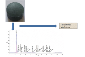
In the present study, S. platensis was cultivated and propagated in a suitable broth medium, then it was available in the form of capsules (Fig. 1). HPLC analysis of S. platensis detected 14 compounds at different retention times. The extract was enriched with certain phenolic and flavonoid compounds including ellagic acid, gallic acid, methyl gallate, naringenin, and chlorogenic acid with different concentrations of 333, 294, 147, 144, and 142 µg/g, respectively. Cinnamic acid, syringic acid, and apigenin were detected in low levels of 2.78, 5.87, and 6.43 µg/g, respectively. Other compounds were recognized in moderate concentrations such as caffeic acid, catechin, and ferulic acid. However, there was a lack of pyro catechol, rutin, coumaric acid, vanillin, and hesperetin in the extract.

Fig. 1. Available form S. platensis in broth (A) and capsule form (B)
Table 1. Flavonoid and Phenolic Compounds Detected in S. platensis Extract

From previous analysis, different phenolic compounds were detected in Spirulina sp., such as gallic, caffeic, salicylic, and trans-cinnamic acids (Pagnussatt et al. 2014). Other studies identified these four compounds besides chlorogenic acid. This difference may occur due to varying conditions of cultivation. The phenolic acid (ellagic acid) in the S. platensis extract in current findings reflected inhibitory action toward aflatoxin B1 production by 82% (Pierzynowska and Grzesiuk 1998). The enhancement of fermentation quality, maintenance of protein, and a decrease in the abundance of dangerous microorganisms in stylo silage have been observed after the addition of ellagic acid (Zou et al. 2021). Exogenous chlorogenic acid exhibited the inhibition of mycotoxin synthesis by Fusarium spp. This result was mainly dependent on the concentration of phenolic acid (Ferruz et al. 2016a). Ferruz et al. (2016b) tested several phenolic acids such as chlorogenic, caffeic, p-coumaric, and ferulic acids against T-2 and HT-2 mycotoxin production by Fusarium sporotrichioides and Fusarium langsethiae. It was determined that F. langsethiae and F. sporotrichioides biomass were not affected by these acids, while T-2 toxin was reduced to 23.1% and 26.5% using chlorogenic and ferulic acids, respectively.

Fig. 2. HPLC chromatograph of S. platensis extract
The identification approaches documented the occurrence of three filamentous fungi on maize silage: Aspergillus flavus, A. niger, and Fusarium sp. Bakri (2021) demonstrated the presence of Aspergillus sp., Fusarium sp., Penicillium, Mucor sp., and Rhizopus sp. on maize silage. The presence of various fungi, particularly mycotoxigenic on maize silage, were considered unattractive characteristics. A. flavus was used to study the efficacy of S. platensis extract on the aflatoxins production. In a recent review study, several biological activities such as anti-oxidants, anti-biofilm, antifungal, and detoxification of mycotoxins were associated with algae and its products (Yadavalli et al. 2023); the data suggests that S. platensis is detoxicant agent for mycotoxins. The level of mycotoxins production was affected by the period of silage incubation and treatment by S. platensis extract. From Table 1, the mean of detected aflatoxin B1, B2, G1, and G2 was 3.14±0.15 ppb, 0.81±0.08 ppb, 0.46±0.05 ppb, and 0.26±0.23 ppb. On the 10th day, it became 2.37±0.33 ppb, 0.59±0.16 ppb, 0.26±0.22 ppb, and 0.21±0.18 ppb when the silage was treated with S. platensis extract. The treated silage showed less content of aflatoxins B1, B2, G1, and G2 compared to untreated silage on the 15th day. It was expected that the aflatoxins in the treated silage on the 15th day would not exceed the aflatoxins on the 10th day. However, the results were opposite from this. It was found that the inhibition percentage of aflatoxins B2 and G2 production was higher on the 15th day (30.61% and 77.77%) than the 10th day (27.16% and 19.23%). The appearance of aflatoxins on the 15th day indicated that the producer fungus growth was not completely inhibited by the S. platensis extract. The current outcomes were in agreement with other studies. For instance, Christ-Ribeiro et al. (2019) showed that the phenolic compounds of Spirulina sp. were effective against fungal growth with 20.2% inhibition as well as ochratoxin A production with 29% inhibition. In previous studies, methanolic extract of S. platensis was applied to suppress the growth and aflatoxin synthesis of A. flavus and A. niger (Pugazhendhi et al. 2015). Synthesis of several F. graminearum mycotoxins including nivalenol, deoxynivalenol, and trichothecene were inhibited by Spirulina sp. extract enriched with gallic acid (Pagnussatt et al. 2014). The benefit of the application of algal macromolecules is its ability to bind mycotoxins without dissociating in the uncontaminated feed. Mycotoxins then go through the digestive system of animals (Yadavalli et al. 2023).
Table 2. Effect of S. platensis Extract on the Aflatoxins Production at 10th Day and 15th Day

*Average of triplicate values of detected mycotoxins and standard deviations
Glucosamine as a biomarker of treated and untreated (control) A. flavus growth by S. platensis extract was estimated at different incubation periods (Fig. 3). S. platensis extract was effective on the glucosamine content of the fungus biomass at all periods of the incubation compared to the untreated fungus with inhibition of 53.7%, 49.2%, 47.8%, 38.5%, and 35.7% on days 3, 6, 9, 12, and 15. Souza et al. (2011) mentioned that the estimation of glucosamine is the best to examine the influence of S. platensis contents on the development of fungal biomass due to its creation. It was linear along the period in the untreated fungus and is affected by phenolic compounds that reflected antifungal activity via inhibiting the production of glucosamine up to 56%. Similar findings were recorded in previous reports using Aspergillus oryzae (Nagel et al. 2001) and Rhizopus oligosporus (Nopharatana et al. 2003)

Fig. 3. Fungal biomass glucosamine at different incubation periods
CONCLUSIONS
- Overall, outcomes indicated that S. platensis contains several flavonoid and phenolic acids.
- S. platensis extract decreases the production of mycotoxins.
- Based on the result of glucosamine detection, S. platensis extract had a repressive effect on A. flavus growth.
ACKNOWLEDGMENTS
Princess Nourah bint Abdulrahman University Researchers Supporting Project number (PNURSP2023R217), Princess Nourah bint Abdulrahman University, Riyadh, Saudi Arabia
Funding
This research was funded by Princess Nourah bint Abdulrahman University Researchers Supporting Project number (PNURSP2023R217), Princess Nourah bint Abdulrahman University, Riyadh, Saudi Arabia
Conflicts of Interest: The authors declare no conflict of interest
REFERENCES CITED
Abdelghany, T. M., Al-Rajhi, A. M. H., Al Abboud, M. A., Alawlaqi, A. Ganash M., Eman A. M. H., and Ahmed S. M. (2018). “Recent advances in green synthesis of silver nanoparticles and their applications: About future directions. A Review,” BioNanoScience 8, 5-16. DOI: 10.1007/s12668-017-0413-3
Abdelghany, T. M., El-Naggar, M. A., Ganash, M. A. (2017). “PCR identification of Aspergillus niger with using natural additives for controlling and detection of malformins and maltoryzine production by HPLC,” BioNanoSci 7, 588-596. DOI: 10.1007/s12668-017-0455-6
Al-Ghanayem, A. A. (2017). “Antimicrobial activity of Spirulina platensis extracts against certain pathogenic bacteria and fungi,” Adv. Biores 8, 96-101. DOI: 10.15515/abr.0976-4585.8.6.96101
Al-Rajhi, A. M. H., Qanash, H., Almuhayawi, M. S., Al Jaouni, S. K., Bakri, M. M., Ganash, M., Salama, H. M., Selim, S., and Abdelghany, T. M. (2022). “Molecular interaction studies and phytochemical characterization of Mentha pulegium L. constituents with multiple biological utilities as antioxidant, antimicrobial, anticancer and anti-hemolytic agents,” Molecules 27, 4824.27(15). DOI: 10.3390/molecules27154824
Anke, H., Kolthoum, I., Zähner, H., and Laatsch, H. (1980). “Metabolic products of microorganisms. 185. The anthraquinones of the Aspergillus glaucus group. I. occurrence,” Arch. Microbiol. 126(3), 223-30. DOI: 10.1007/BF00409924
Bajpai, V. K. (2016). “Antimicrobial bioactive compounds from marine algae: A mini review,” Indian J. Geomar. Sci., 45, 1076-1085.
Bakri, M. M. (2021). “Evaluating the effects of cellulolytic enzymes and Lactobacillus bulgaricus on mycotoxins production and the quality of maize silage,” BioResources 16(4), 8366-8378. DOI: 10.15376/biores.16.4.8366-8378
Bencheikh, A., Mamache, W., Gharzouli, A., Kouachi, A., Khadidja, H., Daichi, M. B., and Rouag, N. (2022). “Evaluation of the spirulina (Arthrospira platensis Gomont) antimicrobial activity,” Turkish Journal of Agriculture- Food Science and Technology 10(10), 2051-2055. DOI: 10.24925/turjaf.v10i10.2051-2055.5307
Christ-Ribeiro, A., Graça, C. S., Kupski, L., Badiale-Furlong, E., and de Souza-Soares, L. A. (2019). “Cytotoxicity, antifungal and anti mycotoxins effects of phenolic compounds from fermented rice bran and Spirulina sp.,” Process Biochemistry 80, 190-196. DOI: 10.1016/j.procbio.2019.02.007
Cohen, Z. (1997). “The chemicals of Spirulina,” in: Spirulina platensis (Arthrospira): Physiology, Cell-biology and Biotechnology,” A. Vonshak (ed.), Taylor and Francis Ltd, London, pp. 175-204. DOI: 10.1590/S1413-70542011000600003
Domsch, K. H., Grams, W., and Anderson, T.-H. (1980). Compendium of Soil Fungi, Academic Press, London.
El-Taher, E. M., El-Ghany, A., and Ashour, M. S. (2012). “Biosecurity for reducing ochratoxin A productivity and their impact on germination and ultrastructures of germinated wheat grains,” Journal of Microbiology, Biotechnology and Food Sciences 2(1), 135-151.
Fayyad, R. J., Nuaman, R. S., Hamdan, N. T., Hameed, R. S., and Sara, A. J. (2020). “Assessment of antagonistic effect of alcoholic extract from cyanophyta (Spirulina platensis) against several human and plant derived pathogenic fungi,” Indian Journal of Forensic Medicine & Toxicology 14(1), 703-709. DOI: 10.37506/ijfmt.v14i1.134
Ferruz, E., Loran, S., Herrera, M., Gimenez, I., Bervis, N., Barcena, C., and Arino, A. (2016a). “Inhibition of Fusarium growth and mycotoxin production in culture medium and in maize kernels by natural phenolic acids,” Journal of Food Protection 79(10), 1753-1758. DOI: 10.3390/toxins9070198
Ferruz, E., Atanasova‐Pénichon, V., Bonnin‐Verdal, M., Marchegay, G., Pinson‐Gadais, L., Ducos, C., Lorán, S., Ariño, A.; Barreau, C., and Richard‐Forget, F. (2016b). “Effects of phenolic acids on the growth and production of T‐2 and HT‐2 toxins by Fusarium langsethiae and F. sporotrichioides,” Molecules 21, 449. DOI: 10.3390/molecules21040449
Hamed, M. A., Abdel Ghany, T. M., Elhussieny, N. I., and Nabih, M. A. (2016). “Exploration of fungal infection in agricultural grains, aflatoxin and zearalenone synthesis under pH stress,” Int. J. Curr. Microbiol. App. Sci 5(4), 1007-1017.
Harini, A. B., and Rajkumar, R. (2022). “Development of sustainable bioproducts from microalgae biomass: Current status and future perspectives,” BioResources 17(4), 7285-7312. DOI: 10.15376/biores.17.4.Harini
Leszczynska, J., Maslowska, J., Owczarek, A., and Ucharska, U. K. (2001). “Determination of aflatoxins in food products by ELISA method,” Czech J. Food Sci. 19, 8-12.
Magnoli, C. E., Astoreca, A. L., Chiacchiera, S. M., and Dalcero, A. M. (2007). “Occurrence of ochratoxin A and ochratoxigenic mycoflora in corn and corn based foods and feeds in some South American countries,” Mycopathologia 163(5), 249. DOI: 10.1007/s11046-007-9005-z
Nagel, F. J. J., Tramper, J., Bakker, M. S., and Rinzema, A. (2001). “Model for on‐line moisture‐content control during solid‐state fermentation,” Biotechnology and Bioengineering 72(2), 231-243. DOI: 10.1002/1097-0290(20000120)
Nopharatana, M., Mitchell, D. A., and Howes, T. (2003). “Use of confocal scanning laser microscopy to measure the concentrations of aerial and penetrative hyphae during growth of Rhizopus oligosporus on a solid surface,” Biotechnology and Bioengineering 84(1), 71-77. DOI: 10.1002/bit.10752
Oltu, I., and Rudic, V. (2016). “Antifungal activity of extracts from Arthrospira platensis against some pathogens, causing invasive mycoses,” Curierul Medical 59(6), 9-14.
Pagnussatt, F. A, Del Ponte, E. M., Garda-Buffon, J., and Badiale-Furlong, E. (2014). “Inhibition of Fusarium graminearum growth and mycotoxin production by phenolic extract from Spirulina sp.,” Pesticide Biochemistry and Physiology 108, 21-26. DOI: 10.1016/j.pestbp.2013.11.002
Penagos-Tabares, F., Khiaosa-Ard, R., Schmidt, M., Pacífico, C., Faas, J., Jenkins, T., Nagl, V., Sulyok, M., Labuda, R., and Zebeli, Q. (2022). “Fungal species and mycotoxins in mouldy spots of grass and maize silages in Austria,” Mycotoxin research 38(2), 117-136. DOI: 10.1007/s12550-022-00453-3
Pierzynowska, J., and Grzesiuk, E. (1998). “Antimutagenic effects of ellagic acid, rutin and psoralen against aflatoxin B1,” Journal of Animal and Feed Sciences 7(Suppl. 1), 277-283. DOI: 10.22358/jafs/69990/1998
Pugazhendhi, A., Margaret, M. R. A., and Mary, S. J. (2015). “Antifungal activity of cell extract of Spirulina platensis against aflatoxin producing Aspergillus species,” International Journal of Current Microbiology and Applied Sciences 4(8), 1025-1029.
Ramamurthy, V., and Raveendran, S. (2009). “Antibacterial and antifungal activity of Spirulina platensis and Lyngbya majuscula,” Journal of Ecobiology 24(1), 47-52.
Raper, K. B., and Fennell, D. I. (1973). The Genus Aspergillus, Lippincott Williams & Wilkins, Philadelphia, PA.
Renganathan, R., Zahira, Y., and Mohd, S. T. (2014). “Potential of the micro and macro algae for biofuel production: A brief review,” BioResources 9(1), 1606-1633. DOI:10.15376/biores.9.1.1606-1633
Salem, O. M., Abdelsalam, A., and Boroujerdi, A. (2021). “Bioremediation potential of Chlorella vulgaris and Nostoc paludosum on azo dyes with analysis of metabolite changes,” Baghdad Science Journal, 18(3), 445-454. DOI: 10.21123/bsj.2021.18.3.0445
Salem, O., Hammad, A. M., Ismail, S., and Elgendy, A. E. M. (2019). “Bio-treatment of wastewater using mixed algal cultures,” Arab Universities Journal of Agricultural Sciences, 27(3), 1871-1880. DOI: 10.21608/AJS.2019.12328.1021
Scotti, C. T., Vergoignan, C., Feron, G., and Durand, A. (2001). “Glucosamine measurement as indirect method for biomass estimation of Cunninghamella elegans grown in solid state cultivation conditions,” Biochemical Engineering Journal 7(1), 1-5. DOI: 10.1016/S1369-703X(00)00090-5
Souza, M. M. D., Prietto, L., Ribeiro, A. C., Souza, T. D. D., and Badiale-Furlong, E. (2011). “Assessment of the antifungal activity of Spirulina platensis phenolic extract against Aspergillus flavus,” Ciência e Agrotecnologia 35(6), 1050-1058.
Yadavalli, R., Valluru, P., Raj, R., Reddy, C. N., and Mishra, B. (2023). “Biological detoxification of mycotoxins: Emphasizing the role of algae,” Algal Research 71, article 103039. DOI: 10.1016/j.algal.2023.103039
Zou, X., Chen, D., Lv, H., Zhang, Q., and Zheng, P. (2021). “Effect of ellagic acid on fermentation quality and bacterial community of stylo silage,” Fermentation 7(4), 256. DOI: 10.3390/fermentation7040256
Article submitted: March 14, 2023; Peer review completed: May 6, 2023; Revised version received and accepted: May 7, 2023; Published: May 12, 2023.
DOI: 10.15376/biores.18.3.4532-4542