
Abstract
The hyper yield of peroxidase (POX) was investigated for a novel native Aspergillus niger strain identified by 18S RNA analysis. A. niger strains sequences were submitted to GenBank; IDs allotted were MN611114.1 (BMB 17) and MN559756.1 (BMB-18). The identified Aspergillus strains in combination showed enhanced (POX) activity (601.5 U/mL) by solid-state fermentation in comparison to their individual activities. POX was purified by ammonium sulfate, and size exclusion gel chromatography exhibited a 7.83-fold increase in POX concentration (13.3 U/mg) in comparison to BMB17 and BMB 18 (11.8 & 7.6 U/mg respectively). The best POX activity was obtained with pH 6.5, 37 °C, and 5 days of incubation. Using guaiacol as substrate, POX showed maximum activity (Vmax) of 537 U/mL with a corresponding Michaelis constant (Km) value of 126 µM. Calcium chloride worked as a POX activator at 300 & 400 mM. Zinc sulfate (500 mM), EDTA (5 mM); ethanol, propanol, and acetonitrile (50%) inhibited (18-30%) POX. Urea (1M), and copper sulfate (500 mM) strongly inhibited POX up to 40%. Polysorbate-80 (1%) slightly reduced the POX by 10% to 15%. BMB17+18-induced promising dye decolorization (88-98%) against all vat dyes, methylene blue, and phenol red.
Download PDF
Full Article
Physicochemical Parameters Optimization and Peroxidase Characterization from Aspergillus niger Native Strain by Solid-State Fermentation for Improved Dye Decolorization
Shiv Ram Ashraf, Amber Afroz,* and Zahid Anwar
The hyper yield of peroxidase (POX) was investigated for a novel native Aspergillus niger strain identified by 18S RNA analysis. A. niger strains sequences were submitted to GenBank; IDs allotted were MN611114.1 (BMB 17) and MN559756.1 (BMB-18). The identified Aspergillus strains in combination showed enhanced (POX) activity (601.5 U/mL) by solid-state fermentation in comparison to their individual activities. POX was purified by ammonium sulfate, and size exclusion gel chromatography exhibited a 7.83-fold increase in POX concentration (13.3 U/mg) in comparison to BMB17 and BMB 18 (11.8 & 7.6 U/mg respectively). The best POX activity was obtained with pH 6.5, 37 °C, and 5 days of incubation. Using guaiacol as substrate, POX showed maximum activity (Vmax) of 537 U/mL with a corresponding Michaelis constant (Km) value of 126 µM. Calcium chloride worked as a POX activator at 300 & 400 mM. Zinc sulfate (500 mM), EDTA (5 mM); ethanol, propanol, and acetonitrile (50%) inhibited (18-30%) POX. Urea (1M), and copper sulfate (500 mM) strongly inhibited POX up to 40%. Polysorbate-80 (1%) slightly reduced the POX by 10% to 15%. BMB17+18-induced promising dye decolorization (88-98%) against all vat dyes, methylene blue, and phenol red.
DOI: 10.15376/biores.18.3.5512-5530
Keywords: Peroxidase; 18S RNA; SSF; Bioremediation; Vat dyes
Contact Information: Department of Biochemistry and Biotechnology, University of Gujrat, Hafiz Hayat Campus Gujrat, Punjab, Pakistan;
* Corresponding author: dramber.afroz@uog.edu.pk; ambernics01@gmail.com
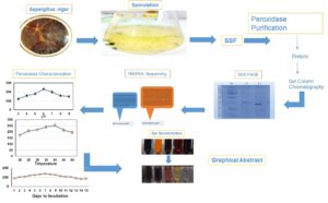
GRAPHICAL ABSTRACT
INTRODUCTION
In third-world countries, the absence of a wastewater treatment plant for industries such as textiles is a big threat. As textile mills produce water that is contaminated with different dyes during dyeing, the highest environmental threat is linked to health (Kishor et al. 2021a). The dyes products and effluents are found to be mutagens and carcinogens, affecting agricultural land, aquatic life, and drinking water (Chang et al. 2021). Currently, wastewater with dye treatment is dependent on chemical management, including redox reactions (reduction, precipitation, filtration, flotation, and ion exchange ozonation). The chemical hazards include expensive large-scale plants and low decolorization efficiency for a variety of dyes in combination (Katheresan et al. 2018). The research group’s focus is to use the fungus and bacterial sources with specific enzymatic potential for dyed effluent treatment. They will have the benefit of using safe biocompatible, with no side effects (Kishor et al. 2021b). Enzymes obtained from a natural sources can act as a biocatalyst, and are the best choice for substrate degradation and the removal of dye (Gurung et al. 2013). The increasing demand for enzymes in bioremediation increases the research demand for their maximum activity (Zamocky et al. 2015). The removal of color from dye-bearing effluent is a problem that conventional methods cannot solve (Adegoke et al. 2015). Different enzymes used for dye decolorization included ligninolytic enzymes. This includes laccase, manganese (Mn) peroxidase, and lignin peroxidase (POX), which were reported for dye and dye decolorization (Liu et al. 2020). POX are grouped into the protoporphyrin IX, cyclooxygenase, and catalase superfamily; these are involved in mammalian, bacterial, and yeast immune responses under oxidative stress by detoxifying reactive oxygen species (Zhuo and Fan 2021; Chen et al. 2023; Zamocky et al. 2015). POX catalyzes the detoxification of substrates with hydrogen peroxide as an electron acceptor that is widely distributed in plants, animals, fungi, bacteria, and mammalian cells (Chen et al. 2023). POX had wide applications as animal fodder, contaminants breakdown, waste-water treatment, dye decolorization, bioethanol production, and for lignin breakdown in pulp and paper industry applications (Bansal and Kanwar 2013; Bilal et al. 2015; Mahmood et al. 2017; Liu et al 2020; Xu et al. 2021). Enhanced POX activity has been reported for Anthracophyllum discolor with optimized media conditions having KH2PO4 and Tris/HCl with a pH of 5.5 at 50 °C (Acevedo et al. 2011). Aspergillus is reported for decolorizing Xiron orange RHD (FW) and scarlet P2R (Kimsa) dyes. It decreased the biochemical oxygen demand of industrial wastewater by 24.2% (Kıvanc and Ozen 2017). In Pakistan, industrialization started in 1960. Approximately 700 factories are registered and approximately the same number are known informally as producing waste in millions of tons (Syed et al. 2010). The industries clustered in Sialkot, Faisalabad, and Lahore are hot-spotted areas with 264 tanneries in Justin Sialkot. These industries produce untreated dye as an effluent (Butt et al. 2021).
Solid-state fermentation (SSF) is an efficient tool for enzyme production using microbes’ culture using small fermenters (Riffat et al. 2022). Lignocellulosic waste (corn cob, wheat straw, and nut shells) degradation in an environmentally friendly mode is critical; especially in underdeveloped countries where the waste management is still inefficient. Corncob is an important agricultural waste: it’s utilization as a POX-producing fungus substrate is cost-effective. Corncob dry biomass comprised of 5 to 36% cellulose, 32 to 40% hemicellulose, 15 to 20% lignin, and 1.0 to 1.7% ash acts as a solid support with great water retention capability: important in fungal growth (Wang et al. 2016; Yu et al. 2021).
A. niger novel strains found to produce Cytochrome C (Cyt), and MnPOX were found promising for dye decolorization. The fungal species were identified by 18S rRNA analysis and compared to other strains by phylogenetic analysis. SSF was used with wheat straw, nutshell, and corncob, as a substrate for enhanced POX production from identified A. niger. In this study, synthetic dyes and POX-producing A. niger samples were collected from areas around tanneries effluents in Sialkot; and the impact on dyes decolorization was explored.
EXPERIMENTAL
Isolation and Characterization of Strains
A. niger from the soil samples near the textile mills in Sialkot, Pakistan was isolated at the Airport Road, Paris Road Cantonment Board Sialkot Pakistan (Fig. 1). Samples were incubated at 37 °C for 5 to 7 days on potato dextrose agar medium (PDA) (King et al. 1986). Pure cultures were maintained at 4 ºC and subsequently subcultured after 30 days. Morphological identification was performed based on colony form (diameter, color of its top, and reverse) while microscopic traits involved conidia and hyphae characteristics (Mohammed 2013).

Fig. 1. Sites of Aspergillus niger (BMB 17, and BMB 18) collection in District Sialkot by GPS Map Camera 1A Site 1, 1B Site 2, 1C Site 3 and 1D Site 4
18SrRNA identification and Polymerase Chain Reaction (PCR)
A. niger mycelium culture was placed in PDA broth at 30 °C for 2 to 3 days for DNA isolation by the CTAB (Doyle and Doyle 1990). The primer sequences were designed by conserved nucleotide sequences from Aspergillus species. Primers were F-5′ GTAGTCATATGCTTGTCTC3′ R-5’TCCGCAGGTTCACCTACGGA3′, by Gene-Link™ USA. The PCR reaction was performed with DNA (20 ng), EF-Taq (SolGent, Korea), 2 μL Taq buffer, 1 μL MgCl2, 0.5 μL dNTP mixture, and 0.5 μL of each forward and reverse primer in a 20 µL reaction mixture. PCR reaction consists of initial denaturation at 95 °C for 2 min followed by 35 cycles of denaturation at 95 °C for 1min. It was followed with annealing (55 °C) and extension (72 °C) for 1 min, with a final extension at 72 °C for 10 min. A partial sequence of 791 and 1300 bps for the 18S rRNA gene was identified.
Phylogenetic Analysis
Sequences obtained were submitted to GenBank. Sequences were used for phylogenetic analysis. A phylogenetic tree was constructed to find out the evolutionary relationship with other Aspergillus and fungal species using the Maximum Likelihood method via Phylogeny.fr (https://www.phylogeny.fr/simple_phylogeny.cgi).
Screening of Isolates for POX Production
The A. niger-identified strains were biochemically identified for POX with Guaiacol and modified PDA synthetic media with glucose (10 g), yeast extract (2 g), NH4NO3 (0.2 g), MgSO4.7H2O (0.5 g), K2HPO4 (1 g), and NaH2PO4.H2O (0.4 g/L) with pH adjusted to 6.5 at 37 oC and 7D for incubation (Bilal et al. 2015).
A. niger Spore Suspension
For the spore suspension (107 to 108 spores/mL), A. niger was cultured for 5 days at 37 ± 1 °C in a medium described above with pH adjusted to 6.5. Basal media preparation was inoculated with fungal spores in-vitro.
POX in the Pre-optimized SSF
SSF was conducted with a 5 g peanut shell, wheat straw, and corn cobs in 100 mL of basal media in a 1 L Erlenmeyer flask. The corn Stover, peanut shell, and wheat straw were obtained from the local area. It was washed, dried (60 °C), ground, and stored for further experimentation. The medium consisted of peanut shell/ wheat straw/corn cobs and was added to KH2PO4 (2 g), MgSO4.7H2O (0.5 g), CaCl2.2H2O (0.1 g), KCl (0.5 g/L), and 5 mL of newly prepared A. niger spores incubated at 30 °C with a pH of 6.5 for 10 days in a static incubator (MIR-254, Sanyo, Japan).
POX Activity
After POX production by A. niger, a flask with spore culture was placed for 10 days under optimum conditions at 37 °C. Extracted enzymes were filtered followed by centrifugation (3000 g) for 10 min at 4 °C. Supernatants were used to check POX activity at 540 nm. POX was detected with basal salts medium containing 0.01% Guaiacol as described (Huy et al. 2017). After, 1D POX was confirmed by oxidized Guaiacol.
Fractional Precipitation for POX Purification
Crude POX broth was subjected to 30 and 50% (NH4)2SO4 saturation for precipitations on ice for 30 min. The mixture was then placed overnight at 4 °C, followed by centrifugation at 5000 g for 30 min. The pellet was dissolved in 50 mM sodium malonic acid buffer (4.1 g malonic acid, 1.83 sodium malonate/L, and a pH of 4.5) and dialyzed against distilled water after 6 h to remove leftover ammonium sulfate (Riffat et al. 2022).
Gel Filtration Chromatography
Sephadex G-100 column (Sigma, USA) column (120x2cm) was used for activated POX purification. Phosphate buffer (100 mM) with 0.15 to 1 M NaCl was used as elution buffer: at pH 6.5 and a flow rate of 0.28 mL per min. Twenty-five active fractions were obtained and checked for maximum POX activity (Goyal and Chugh 2014).
Sodium Dodecyl Sulphate-Polyacrylamide Gel Electrophoresis (SDS-PAGE)
SDS-PAGE was performed using Mini Vertical Gel Electrophoresis apparatus MV-10D SYS (Major Science UK) (W x L=3.4″x3.2″) on 12.5% separating and 5% stacking gel. Extracted protein samples were applied to the gels, along with a protein ladder (Thermo Scientific™ Ladder Cat #: 26616). The gel was then run at 80 V. Gel was stained by Coomassie Brilliant Blue for 2h, followed by destaining. Gel was imaged on a densitometer (GS-900TM Densitometer with Image LabTM Software Version 5.1).
PCR Amplification of POX Gene
A. niger (50 mg) RNA was extracted by RNA extraction kit (PureLink™ RNA Mini Kit: Cat #: 12183020). It was followed by the cDNA synthesis by Kit (Revert Aid First Strand cDNA Synthesis Kit: Cat #: K1622). Primers were designed from NCBI (XM_001401736.2; Cyt POX: 1734 bps; OM456997.1; MnPOX: 1314 bps) by Primer3 and properties were confirmed through the oligonucleotide properties calculator. Primers were used in 5′-3′ direction for Cyt POX: F-ACGGCGCCAGAAATGCG, and R-TGAATCTTCAAAAGGATGGTT; and for MnPOX: F-GTTCTACTTCCTCCTCCT-CCG, and R-GACAGAGATGAACCTCATGTA by Gene-Link™ USA. The reverse transcriptase-polymerase chain reaction (RT-PCR) reaction mixture contains cDNA (1.5 μL), Taq buffer (2 μL), MgCl2 (1 μL), dNTP (0.5 μL), forward and reverse primer (0.5 μL), Taq polymerase (1 μL), and Nano pure water for a volume up to 20 μL. PCR cycle involved denaturation at 95 °C (4 min) followed by 40 cycles of denaturation at 95 oC (30 s). The annealing temperature was 55 °C, and 57 °C (1.5 min), for Cyt, and MnPOX respectively, followed by the extension at 72 °C (30 s), with a final extension at 7 min. The amplified product was run on 1% agarose gel and Photographed on a gel documentation system.
Characterization of Purified POX
The A. niger (BMB17, BMB18, and BMB17+18) showed the highest POX activity and was further optimized for its enhanced production. The optimized POX production was determined with changing moisture content (10 to 90%), ammonium sulfate (1 to 5.5 mg mL-1), MgSO4 (0.2 to 0.8 mgL-1), and KH2PO4 (0.1 to 0.6 mgL-1), along with temperature and incubation period at 24 h intervals.
POX Characterization
POX activity was determined with a change in pH (2.5 to 8.5), temperature range of 25 to 52 °C, and days to incubation (1 to 11 D) with a combination of buffers at optimized KH2PO4, NH4SO4, and MgSO4 (Fig. 3A, 3B, 3C).
Determination of Vmax and Km
POX kinetic parameters, including the maximum rate of its activity (Vmax) and Michaelis-Menten constant (Km), were determined by plotting a graph of the substrate amount linked to the maximum enzyme activity. The enzyme activity value (UmL-1) was taken at 540 nm in triplicate by examining the 1 to 5 µM substrate (Guaiacol). The reciprocal plot was constructed by taking the reaction rate versus substrate concentration using the Lineweaver-Burk plot transformation of the Michaelis-Menten equation (Fig. 6).
Effect of Activators and Inhibitors
The effect of metal bivalent ions (Ca2+, Cu2+, and Zn2+: 100 to 500 mM) in the form of CaCl2, CuSO4, and ZnSO4 on POX activity was measured. Along with this effect of denaturants (urea: 1 M), chelating agent: EDTA (5 mM), surfactant: Tween 80 (Polysorbate 80) (1%), detergent (SDS 1%), and organic solvents: Ethanol, Acetonitrile, and propanol (50%) were used. The control consists of 0.1 mL crude enzyme, 0.05 mL of 30% H2O2, 0.05 mL of 0.018 M guaiacol, 2.8 mL of 0.1M phosphate buffer (pH 6.5), at 25 oC for 5-6 min with different activators, and inhibitors; optical density was calculated at 595 nm (Huy et al. 2017).
Dye Decolorization
Percentage decolorization was calculated by measuring absorbance at a range of temperatures containing the dyes methylene blue, vat orange, vat black, phenol red, and vat grey. The reaction mixture consisted of 0.05 M acetate buffer (2 mL at pH 5.0), dye (100 µL), H2O2 (100 µL), and POX (100 µL), CaCl2 (300 mM: 200 µL) was incubated at 37 oC for 60 min. The oxidation monitoring was measured 2 h post-incubation of the enzyme by taking absorbance at 665, 430, 630, 460, and 614 λ for Methylene Blue, Vat orange, Vat Black, phenol red, and Vat grey.
The percentage of decolorization was calculated as follows,
% decolorization = (Ai – Af /Ai) x 100 (1)
where Ai is the initial absorbance of untreated dye, and Af is the final absorbance of treated dye.
Statistical Analysis
Microsoft Excel 2013 and Minitab 17 were used for the data analysis and graphs. The POX activity using corn cob, nutshell, and wheat straw as substrates was compared after calculating the means followed by standard deviation (STDEV). The calculations considered different factors affecting POX activity, along with modulators and repressors. One-way analysis of variance (ANOVA) was carried out to find the significant difference in the POX activity against different factors.
RESULTS AND DISCUSSION
POX alternate sources needed to be explored, as its requirement cannot be fulfilled by the main commercial horseradish POX. Fungal POX such as yeast Cyt, lignin, Mn, and versatile POX are involved in heme POX activity, lignin degradation, and ROS scavenging (Zhuo and Fan, 2021; Chen et al. 2023). Biodegradation of organic pollutants in wastewater treatment operations often involves the Fenton reaction. Advanced oxidative products, such as the Fenton reaction, are mediated by the quinone redox cycling of the fungal peroxidases (Chen et al. 2023). White rot fungi (WRF) (Basidiomycetes) can degrade lignin within the wood, giving rise to a bleached look (Bilal et al. 2017). The ligninolytic systems of WRF comprise several major extracellular components, including dye-decolorizing POX, lignin, Mn, versatile peroxidase, Cyt POX, and laccase (Chen et al. 2023; Zhuo and Fan 2021). A. niger (Ascomycetes) peroxidase was reported as being effective for Congo red decolorization (97%). Maximum decolorization of dye (200 mg L-1) was obtained at pH 5, after 6 days of incubation at 28 °C (Asses et al. 2018). Most of the MnP-assisted decolorization had used pure cultures; effects were not as good as those of microbial consortium. A fungal consortium using two microbial consortia (Bacillaceae, Piscibacillus, and Bacillus) and (Halomonas, Marinobacter, and Clostridiisalibacter), resulted in 93% decolorization of 100 mg/L Metanil Yellow G within 48 h. In addition, both consortia were highly effective in high-pH and high-salinity environments, indicating potential use for the treatment of high-salinity and alkaline textile wastewater (Guo et al. 2020). In this study consortium of Cyt, and MnPOX resulted in promising decolorization rather than independently (Table 1; Fig. 4).
POX Production
In this study, enhanced POX production was obtained with use of native fungal strains (BMB17, BMB18, and BMB17+18) through SSF of corn cob with pre-optimized parameters. More POX activity (538 U/mL) was obtained with corn cob inoculation with freshly prepared A. niger BMB-17 (Cyt POX) at 37 °C, pH (6.5), and 5D of incubation. BMB18 (MnPOX) showed 381 U/mL activity of POX, but when effects of both BMB17 and BMB18 (Cyt+ MnPOX) were combined, the POX activity was increased to 602 U/mL (Table 1). Laccase activity (260 U/mL) by Fomes fomentarius after 10D of incubation at 35 °C using corn Stover as a substrate was reported (Riffat et al. 2022). POX activity of 1.049 U at pH 5 at 30 oC was reported (Govarthanan et al. 2017). The 143.9 U POX activity at 60 oC and 4.5 pH were reported (Zhang et al. 2018). Similarly, Jarvinen et al. (2012) reported the 52 U activity of MnPOX at 4.5 pH.
Table 1. Peroxidase Purification by Solid-State Fermentation by Aspergillus niger strains BMB-17, 18, and a combination of 17+18

* Specific Activity = Total activity/Protein conc (U/mg); Purification Fold = Specific activity of Purified Enzyme/Crude Enzyme; Yield (%) = Total U of Purified enzyme/crude enzymex100.
A. niger Identified Strains Phylogenetic Analysis
The black and sulfur-yellow colonies at the top and bottom showed the presence of A. niger. This identification was confirmed by biochemical characterization. There was a brown oxidation zone in 2 fungal strains: Asp1 and Asp2. 18S rRNA A. niger strain was submitted to NCBI and accession numbers (MN611114.1 (BMB17) and MN559756.1 (BMB18) were obtained. Phylogenetic analysis of respective strains shows their alignment with A. niger, A. fumigatus, A. awamori, and A. luchuensis respectively (>95%) (Fig. 2).

Fig. 2. Phylogenetic tree of MN611114.1 (BMB-17) (A) and MN559756.1 (BMB-18) (B). The phylogenetic tree was constructed on phylogeny.fr software after all the closely related Aspergillus and other fungal species BLAST
POX Production and Purification
The selected fungal strain was grown on PDA slants for 5D at 37 °C and stored at 4 °C. Corn cob was used for SSF fermentation, as it provides better than wheat straw and peanut shell. It is composed of 5 to 36% cellulose, 32 to 40% hemicellulose, 15 to 20% lignin, and 1.0 to 1.7% ash (Chang et al. 2021) with water retention and solidity indicating fungus cultivation potential. BMB17+18 SSF shows POX activity of 601.5 U/mL.
POX purification with (NH4)2SO4 fractionation with specific and total activity of 3.85, 2.3, and 4.12 U/mg by BMB 17, 18, and a combination of 17 and 18 (Table 1). Sephadex G-100 gel filtration column with maximum and specific POX activity (150.34 and 13.3 U/mL) was obtained and purification augmented 7.8 folds with BMB17 and 18 (Table 1). An earlier study by Goyal and Chugh (2014) reported Pennisetum glaucum POX purification (19%) with 24 mg/39 mL recovery by size exclusion chromatography. The 12.5% SDS PAGE shows a 40 kDa POX band with standard molecular weight markers (Fig. 3A). The PCR showed a POX with a band size of 1734, and 1314 bps from cDNA (Fig. 3B; C).

Fig. 3. (A) SDS PAGE of purified peroxidase from A. niger strain. M (Bio-Rad SDS Ladder) (KDa) L1 (BMB17), L2 (BMB18); (B) Amplification of peroxidase from A. niger BMB17: peroxidase gene (1734 bps), (M) 1Kb Ladder (L2) Cyt POX; (C) Amplification of peroxidase from A. niger BMB18: Mn Peroxidase gene (1314 bps), (M) 1Kb Ladder (L1) MnPOX
The highest laccase activity of 1.479 U/mL was reported by El Monssef et al. (2016); whereas the laccase and azoreductase initial activity of 3.2 U/mL from the liquid culture medium by the Aeromonas hydrophila, was reported against textile dyes (Srinivasan et al. 2019).
Characterization
The study optimized extracellular POX BMB17, BMB18, and BMB17+18 by substrate optimization (pH, temperature, and incubation period) (Xu et al. 2021). Peanut shells, wheat bran, corn cobs, and sukhchain shells plant lignocellulosic materials were reported as a source of SSF (Oliaei et al. 2021). In this experiment, corn cob was optimum for maximum POX activity. Values of 280, 320, and 350 U/mL activity were recorded for nutshell, wheat straw, and Corn cob respectively after 7D of incubation (pH 6.5, 37 °C) (Fig. 4).
Moisture (10%), MgSO4 (0.6 g), (NH4)2SO4 (4 g), KH2PO4 (0.5 g), and substrate concentration (1 mgmL-1) were optimum for maximum enzyme activity (391.6 U/mL). POX activity of 1,354 U/L was reported with 50 mM KH2PO4, Tris/HCl, pH 5.5, at 50 ºC (Acevedo et al. 2011). Falade et al. (2019) reported K2HPO4 (4.5 g), KH2PO4 (0.53 g), MgSO4 (0.5 g), NH4NO3 (5 g), yeast extract (0.1 g), 0.1% w/v alkali lignin/L for bacterial POX. Ammonium sulfate and size exclusion chromatography purification resulted in 2.4 and 7.8-fold increased POX activity respectively (Table 1). The 1734, and 1314 bps, with 40 KDa protein bands confirmed Cyt, and Mn POX, respectively (Fig. 3).

Fig. 4. Effect of corn cob, wheat straw, and peanut shell on peroxidase 1-11D post-incubation. Maximum activity was observed by corn cob 7D-post incubation by SSF
Effects of pH value, Temperature, and incubation days on POX Activity
Optimum POX activity was obtained at pH 6.5 (235 U/mL) (BMB17+18=390 in comparison to 379.23 and 270 for BMB 17 and 18), 37 °C (BMB17+18=423.2 in comparison to 330 and 290 for BMB 17 and 18), and 5D incubation (BMB17+18=537 in comparison to 432.5 and 400 for BMB 17 and 18) (Fig. 5A, B, C). The pH of the medium impacts the microbe’s net charge, affecting its enzymatic production physiology (Makapela et al. 2016). Turnip POX at pH 8 and 30 °C gave maximum activity (4027 U/mg) (Dahdouh et al. 2020). Falade et al. (2019) reported 5.9 pH at 30 °C in Ensifer adhaerens POX. Musengi et al. (2014) reported 37 °C for Bacillus subtilis and Streptomyces sp. POX. Optimized temperature variation caused metabolic activities to decrease, leading to enzyme active site modification (Saez et al. 2019).

Fig. 5. Characterization and optimization of different parameters on the A. niger peroxidase activity (A) pH (B) Temperature (C) Days to incubation
Enzyme Kinetics and POX Production
Purified POX Vmax and Km were calculated with the concentration substrate range for a reciprocal graph (Fig. 6). The purified enzyme Km value was 126 µM with a reaction velocity of 537 UmL-1 with the substrate (guaiacol). A. niger POX with H2O2 and guaiacol as a substrate had values of Km obtained which were 0.751 mM for Li and Mn POX while Vmax values were 1000 to 1250 µM/mL (Mahmood et al. 2017).

Fig. 6. The reciprocal plot of 1/[S] (µM) and 1/[V] (UmL-1) for determination of Michaelis constant (Km) in µM and Vmax UmL-1 of Aspergillus niger BMB17+18 peroxidase
Similarly, the value of purified Trametes versicolor laccase Km of 73 µM and 780 U/mL Vmax was obtained using 2,2′-azinobis [3-ethylbenzothiazoline-6-sulfonic acid]-diammonium salt as substrate (Asgher et al. 2012). Ginger POX exhibited Km values of 7.1 mM and Vmax values of 0.31 U/assay using guaiacol as substrate (El-Khonezy et al. 2020). Humicola grisea POX represents Km values of 1.25 to 5.56 μM for different isozymes, while the Vmax values were 0.45 to 0.6 U/mg (Moubasher et al. 2017). Ganoderma lucidum Mn POX Km and Vmax with MnSO4 as a substrate were 65.5 mM and 640 UmL-1 (Bilal et al. 2015). Relatively lower Km and high Vmax values compared to previous reports support the high substrate affinity for the enzyme (Goyal and Chugh 2014). POX was optimized by SSF and the concentration obtained was 1.7 U/g enhanced by ammonium sulfate precipitation to 3.04 U/mg (Table 1). Gel filtration chromatography resulted in the purification of POX by 7.83 folds with an increase in specific enzymatic activity of 13.3 U/mg (Table 1).
Effect of Activators and Inhibitors
Calcium chloride worked as an activator for POX with maximum activity found at 300 mM. CuSO4 and ZnSO4 were moderate inhibitors at higher concentration from 200 to 500 mM (Fig. 7A, B, C). For the binding between dyes and fabric, especially for vat dyes, different chemicals such as metals, salts, and surfactants (detergents: improve solubility), sulfide, and formaldehyde are used (Chang et al. 2021; Kishor et al. 2021 b). Therefore, the effect of the detergents, surfactants, and reducing agents needed to be identified.
Kouakou et al. (2009) reported POX maximum activity at 25 °C with metal ions: Al3+, Fe3+, Ca2+, and Ni2+ while moderately inhibited by Mn2+ and K+. POX lost 50 to 62% of its activity with Zn2+ and Hg2+. Iron, magnesium, and copper in higher concentrations can enhance the oxidative site of the guaiacol substrate (Moubasher et al. 2017). Cu+2 also acts as an activator from Ganoderma lucidum IBL-05 POX (Bilal and Asgher 2015). Copper sulphate inhibits the POX to 40% at 500 mM. Studies are consistent with Ganoderma leucocontextum laccase where Cu+2 acts as an inhibitor (Umar and Ahmed 2022).

Fig. 7. Effect of (A) CaCl2 (B) CuSO4 (C) ZnSO4 (D) Denaturants (Urea), chelating agent: EDTA, Detergent (SDS), Surfactant (polysorbate 80), and Organic solvent (Ethanol, Propanol, Acetonitrile) on peroxidase activity
The EDTA concentration range in dye decolorization experiments was 0.1 to 10 mM; this was found to be effective in chelating metal ions and stabilizing POX activity without excessively inhibiting enzyme function. EDTA could be made POX inactivate when used in high concentration (Duan et al. 2018). EDTA with POX for dye decolorization involves an interplay between the enzyme, the dye, and the chelating agent, leading to improved strategies for efficient and reliable dye removal. It may increase the stability and longevity of POX and optimize the decolorization of dye (Asif et al. 2017). Duan et al. (2018) reported 0.1 mM EDTA as a modulator and 7 to 8 mM EDTA as an inhibitor of MnPOX. The authors have reported 5 mM EDTA as a repressor. Bilal et al. (2015) reported the inhibition of MnPOX by EDTA, cysteine, and Hg2+. Meanwhile, EDTA and ZnSO4 slightly reduced the POX to 20 to 30% (Fig. 7C, D).
Ca+2 was required for the stabilization of the heme in the active site of POX (Moubasher et al. 2017). In this case the calcium ion acted as an activator at higher concentrations like 400 mM, while copper and zinc slightly reduced the POX at higher concentrations of 200 to 500 mM (Fig. 7). The present results showed that calcium activates the POX to 150% at 400 mM while becoming normal at 500 mM (Fig. 7A). The calcium ions are essential components of MnP that provide thermal stability. The other factors reducing the POX activity are concerned with the decrease in oxidizing potential by decreasing the oxidation of POX substrates (Asgher et al. 2012). Studies are consistent with Irpex lacteus POX activated by Ca2+. Oppositely white rot fungus Trametes sp. POX is not affected by Ca2+ and Zn2+ (Qin et al. 2014; Zhang et al. 2016). The organic solvents also help in the solubilization and stabilization of dyes with POX. Copper, calcium, and zinc ions at 10 mM decreased the POX to 10% or remain the same (Lueangjaroenkit et al. 2019; Riffat et al. 2022).
From 0 to 2 M urea was tested: 1 to 2 M urea was found inhibitory. 1 M urea was used for the comparison of 2 POX classes (Fig. 7). Lueangjaroenkit et al. (2019) reported non-ionic surfactants such as polysorbate 80 to be more inhibitory than polysorbate 20 (1-5%) for MnPOX. Here also a slight inhibition was recorded for polysorbate 80 (1%) (Fig. 6). The effect of organic solvents such as ethanol and propanol (50%) was found inhibitory in MnPOX (70, and 43%), while a consortium of Cyt and MnPOX slightly inhibited (30, & 20%) (Fig. 7).
Detergent SDS slightly decreased POX being an important part of textile effluents (Fig. 7D). Conversely, SDS was a strong inhibitor for the laccase activity compared to EDTA at all concentrations (Umar and Ahmed 2022). Polysorbate-20 and EDTA were found to act as inhibitors of Trametes polyzona KU-RNW027 POX (Zhang et al. 2016).
Alcohol at 50% slightly reduced the POX. However, at higher concentrations, POX was found to be inhibitory below 37 oC (Fig. 7D). Trametes polyzona KU-RNW027 MnPOX was activated by methanol, ethanol, propanol, isopropanol, and acetone (Lueangjaroenkit et al. 2019).
Dye Decolorization
The use of A. niger with combined BMB 17+18 POX for maximum dye decolorization had been identified. The main dyes used in textiles are direct, basic, disperse, reactive, pigment, and vat. Textile dyes, namely methylene blue, Vat orange 2, Vat Black 27, phenol red, and Vat Grey 23 at the given wavelength, were analyzed for decolorization with a spectrophotometer after enzyme incubation. Changes in the optical density were recorded after 2 h dye incubation (Fig. 8; Table 2). The maximum decolorization was found in all vat dyes, methylene blue, and phenol red. The decolorization 90.3, 98.2, 88, 93.2, and 88.3% in methylene blue, Vat Orange 2, Vat Black 27, phenol red, and Vat Grey 23 (Fig. 8; Table 2). Enzymatic dye decolorization had advantages over physical, biological, and chemical processes over a wide range of pH, salinity, and simplicity of the controlled process (Kishor et al. 2021a).
Table 2. Dye Decolorization Percentage Calculated by the Optical Density Difference (initial absorbance minus the final absorbance of the treated dyes) by Peroxidase Sources: Aspergillus niger Strains BMB17, 18, and17+18


Fig. 8. Dye Decolorization percentage: Calculated by the optical density difference (initial absorbance minus the final Absorbance of the treated dyes) by peroxidase obtained from the sources: Aspergillus niger strains BMB17, 18, and17+18.
Zhang et al. (2016) reported the use of purified POX from white rot fungus (100 mgL-1) for the removal of synthetic dyes with 85-94.6% efficiency within 2 h. A. fumigatus degraded azo dye with decolorization ranging from 71 to 93% (Abd El-Rahim et al. 2017). Omar (2016) reported A. niger POX’s role in decolorizing textile reactive dyes such as azo, and anthraquinone at a pH of 4. Asgher et al. (2008) reported 74 to 90% of vat dyes decolorization by Coriolus versicolor laccase in 7 days. Zhang et al. (2018) reported the azo dyes decolorization by POX in the range of (53.9 to 81%), and 62% for bromophenol blue in 12 h. Vat dyes are a special synthetic dye insoluble class based on indigo (a natural dye) that needs a reducing agent to solubilize (Katheresan et al. 2018; Chang et al. 2021). The present study successfully decolorized the vat dyes along with others within 2 hours with native Cyt and MnPOX.
Bilal et al. (2015) reported MnPOX from G. lucidum against different synthetic dyes up to (67 to 81%) within 12 hours of incubation. Liu et al. (2020) reported 35-40% decolorization against azodyes; while 65% against bromophenol blue. But it was not effective against mordant yellow and disperse blue with recombinant Aspergillus POX. Daedalea dickinsii MnPOX with maximum activity (612.31 U/mL) can degrade 70 to 80% while Piptoporus betulinus can degrade 47 to 59% textile dyes (Mahmood et al. 2017). The POX caused dye degradation because of active free radicals including lithium, Mn, lipid, hydroxyl, and peroxy-radicals. However, they did not biodegrade equally because of their variation in chemical structures (Mahmood et al. 2017; Liu et al. 2020). SDS is a strong anionic detergent, and polysorbate 80 as a surfactant solubilizes hydrophobic substances, including dyes. It forms micelles along with urea in aqueous solutions, creating a hydrophobic environment within the micelle core where hydrophobic dyes are dissolved. SDS can somehow help to stabilize the POX as well enhance its decolorization ability. Calcium also stabilized POX by interacting with negatively charged amino acids on POX forming stable ionic interactions (Asif et al. 2017; Lueangjaroenkit et al. 2019; Chang et al. 2021; Chen et al. 2023).
CONCLUSIONS
- A. niger strains were identified (MN611114.1 (BMB 17); MN559756.1 (BMB-18), for Cyt and MnPOX with its activity enhanced to 7.29-folds with chromatographic purification. The combined effect of Cyt+ Mn POX activity obtained was 601 U/mL.
- The maximum dye decolorization was found in all vat dyes, methylene blue, and phenol red with a consortium of BMB 17, 18. Ca2+ act as a stabilizer for dye decolorization at a higher concentration such as 300, and 400 mM. The metal ions, organic solvents at higher concentrations do not interfere much with the POX activity. All these factors impart Cyt+ MnPOX as a strong decolorizing agent in wastewater management. The results are not affected by metal ions, detergents, and surfactants.
ACKNOWLEDGEMENTS
Funding
Amber Afroz and Zahid Anwar received research grants from the Higher Education Commission, and the National Research Program of Universities (Grant numbers: NRPU6506 and NRPU4555 respectively).
Declaration
The authors declare that the paper sent is original and no part had been published before or is being considered for publication in any other journal.
Authors’ Contributions
Shiv Ram Ashraf: Experimental Execution, Initial draft write-up; Amber Afroz: Supervision, Sources, Experimental design, Zahid Anwar: Co-Supervision, Experimental design, Technical review, Validity.
REFERENCES CITED
Abd El-Rahim, W. M., Moawad, H., Azeiz, A. Z. A., and Sadowsky, M. J. (2017). “Optimization of conditions for decolorization of azo-based textile dyes by multiple fungal species,” Journal of Biotechnology 260, 11-17. DOI: 10.1016/j.jbiotec.2017.08.022
Acevedo, F., Pizzul, L., Castillo, M. D. P., Rubilar, O., and Lienqueo, M. E. (2011). “A Practical culture technique for enhanced production of manganese peroxidase by Anthracophyllum discolor Sp4,” Brazilian Archives Biology Technology 54(6), 1175-1186. DOI: 10.1590/s1516-89132011000600013
Adegoke, K. A., and Bello, O. S. (2015). “Dye sequestration using agricultural wastes as adsorbents,” Water Resources and Industry 12, 8-24. DOI: 10.1016/j.wri.2015.09.002
Asgher, M., Iqbal, H. M. N., and Asad, M. J. (2012). “Kinetic characterization of purified laccase produced from Trametes versicolor IBL-04 in solid state bio-processing of corncobs,” BioResources 7(1), 1171-1188
Asgher, M., Batool, S., Bhatti, H. N., Noreen, R., Rahman, S. U., and Asad, M., J. (2008). “Laccase mediated decolorization of vat dyes by Coriolus versicolor IBL-04,” International Biodeterioration and Biodegradation 62, 465-470. DOI: 10.1016/j.ibiod.2008.05.003
Asses, N., Ayed, L., Hkiri, N., and Hamdi, M. (2018). “Congo red decolorization and detoxification by Aspergillus niger: Removal mechanisms and dye degradation pathway,” BioMed Research International, article ID 3049686. DOI: 10.1155/2018/3049686
Asif, M. B., Hai, F. I., Hou, J., Price, W. E., and Nghiem, L. D. (2017). “Impact of wastewater derived dissolved interfering compounds on growth, enzymatic activity and trace organic contaminant removal of white rot fungi–a critical review,” Journal of Environmental Management 201, 89-109
Bansal, N., and Kanwar, S. S. (2013). “Peroxidase(s) in environment protection,” The Scientific World Journal 714639, 1-9. DOI: 10.1155/2013/714639.
Bilal, M., and Asgher, M. (2015). “Dye decolorization and detoxification potential of Ca-alginate beads immobilized manganese peroxidase,” BMC Biotechnology 15(1), 1-14. DOI: 10.1186/s12896-015-0227-8
Bilal, M., Asgher, M., and Ramzan, M. (2015). “Purification and biochemical characterization of extracellular manganese peroxidase from Ganoderma lucidum IBL-05 and its application,” Scientific Research and Essays 10(14), 456-464. DOI: 10.5897/SRE2015.6268
Butt, M. Q., Zeeshan, N., Ashraf, N. M., Akhtar, M. A., Ashraf, H., Afroz, A., and Naz, S. (2021). “Environmental impact and diversity of protease-producing bacteria in areas of leather tannery effluents of Sialkot, Pakistan,” Environmental Science and Pollution Research 28(39), 54842-54851
Chang, Y., Yang, D., Li, R., Wang, T., and Zhu, Y. (2021). “Textile dye biodecolorization by manganese peroxidase,” A Review Molecules 26, article 4403. DOI: 10.3390/molecules261544033
Chen, S., Zhu, M., Guo, X., Yang, B., and Zhuo, R. (2023). “Coupling of Fenton reaction and white rot fungi for the degradation of organic pollutants,” Ecotoxicology and Environmental Safety 254, article 114697. DOI: 10.1016/j.ecoenv.2023.114697. PMID: 36889210
Dahdouh, A., Bachir-bey, M., and Kati, D. E. (2020). “Optimization of peroxidase activity of turnip (Brassica rapa) using response surface methodology,” Acta University Cibiniensis Ser E: Food Technology 24(2), 186-194. DOI: 10.2478/aucft-2020-0017
Doyle, J. J., and Doyle, J. L. (1990). “Isolation of plant DNA from fresh tissue,” Focus 8(12), 13-15.
Duan, Z, Shen, R., Liu, B., Yao, M., and Jia, R. (2018). “Comprehensive investigation of a dye-decolorizing peroxidase and a manganese peroxidase from Irpex lacteus F17, a lignin-degrading basidiomycete,” AMB Express, 8(1), 119. DOI: 10.1186/s13568-018-0648-6
El-Khonezy, M. I., Abd-Elaziz, A. M., Dondeti, M. F., Fahmy, A. S., and Mohamed, S. A. (2020). “Purification and characterization of cationic peroxidase from ginger (Zingiber officinale),” Bulletin of the National Research Centre, 44(1), 1-9. DOI: 10.1186/s42269-019-0264-x
Falade, A., Jaouani, A., Mabinya, L., Okoh, A., and Nwodo, U. (2019). “Exoproduction and molecular characterization of peroxidase from Ensifer adhaerens,” Applied Science 9, article 3121. DOI: 10.3390/app9153121
Govarthanan, M., Fuzisawa, S., Hosogai, T., and Chang, Y. C. (2017). “Biodegradation of aliphatic and aromatic hydrocarbons using the filamentous fungus Penicillium sp. CHY-2 and characterization of its manganese peroxidase activity,” RSC Advances 7(34), 20716-20723.
Goyal, P., and Chugh, L. K., (2014). “Partial purification and characterization of peroxidase from pearl Millet (Pennisetum glaucum [L.] R. Br.) grains,” Journal of Food Biochemistry 38(2), 150-158. DOI: 10.1111/jfbc.12033
Guo, G., Hao, J., Tian, F., Liu, C., Ding, K., Zhang, C., Yang, F., and Xu, J. (2020). “Decolorization of Metanil Yellow G by a halophilic alkalithermophilic bacterial consortium,” Bioresour. Technol. 316, article 123923
Guo, G., Hao, J., Tian, F., Liu, C., Ding, K., Xu, J., Zhou, W., and Guan, Z., (2020). “Decolorization and detoxification of azo dye by halo alkaliphilic bacterial consortium: Systematic investigations of performance, pathway and metagenome,” Ecotoxicol. Environ. Saf. 204, article 111073.
Gurung, N., Ray, S., Bose, S., and Rai, V. A. (2013). “Broader view: Microbial enzymes and their relevance in industries, medicine, and beyond,” BioMed Res. Int. 2013: article 329121. DOI: 10.1155/2013/329121
Huy, N. D., Tien, N. T. T., Huyen, L. T., Quang, H. T., Tung, T. Q, Luong, N. N., and Park, S. M. (2017). “Screening and production of manganese peroxidase from fusarium sp. on residue materials,” Mycobiology 45(1), 52-56. DOI: 10.5941/MYCO.2017.45.1.52
Jarvinen, J., Taskila, S., Isomäki, R., and Ojamo, H. (2012). “Screening of white-rot fungi manganese peroxidases: a comparison between the specific activities of the enzyme from different native producers,” Amb Express 2, 1-9.
Katheresan, V., Kansedo, J., and Lau, S. Y. (2018). “Efficiency of various recent wastewater dye removal methods. A review,” Journal of Environmental Chemical Engineering 6, 4676-4697. DOI: 10.1016/j.jece.2018.06.060
King, A. D., Bitt, J. I., Beuchat, L. R., and Corry, J. E. L. (1986). in: Methods for Mycological Examination of Foods, Plenum Press, New York and London 122,315. DOI: 10.1007/978-1-4684-8453-3
Kishor, R., Purchase, D., Saratale, G. D., Saratale, R. G., Ferreira, L. F. R., Bilal, M., Chandra, R., and Bharagava, R. N. (2021a). “Ecotoxicological and health concerns of persistent coloring pollutants of textile industry wastewater and treatment approaches for environmental safety,” Journal of Environmental Chemical Engineering 9, article 105012. DOI: 10.1016/j.jece.2020.105012
Kishor, R., Saratale, G. D., Saratale, R. G., Ferreira, L. F. R., Bilal, M., Iqbal, H. M., and Bharagava, R. N., (2021b). “Efficient degradation and detoxification of methylene blue dye by a newly isolated ligninolytic enzyme producing bacterium Bacillus albus MW407057,” Colloids Surf. B Biointerfaces 206, article 111947. DOI: 10.1016/j.colsurfb.2021.111947
Kıvanc, M., and Ozen, M. D. (2017). “Screening of fungi for decolorization of dye wastewater,” International Proceedings of Chemical, Biological and Environmental Engineering (IPCBEE) 100: 1-7. DOI: 10.1016/j.copbio.2011.05.211
Kouakou, T. H., Due, E. A., Kouadio, N. E. J. P., Niamke, S., Kouadio, Y. J., and Merillon, J. M. (2009). “Purification and characterization of cell suspensions peroxidase from cotton (Gossypium hirsutum L.),” Applied Biochemistry and Biotechnology 157(3), 575-592. DOI: 10.1007/s12010-008-8287-z
Liu, S., Xu, X., Kang, Y., Xiao, Y., and Liu, H. (2020). “Degradation and detoxification of azo dyes with recombinant ligninolytic enzymes from Aspergillus sp. with secretory overexpression in Pichia pastoris,” R Soc Open Sci. 7(9), article 200688. DOI: 10.1098/rsos.200688
Lueangjaroenkit, P., Teerapatsakul, C., Sakka, K., Sakka, M., Kimura, T., Kunitake, E., and Chitradon, L. (2019). “Two manganese peroxidases and a laccase of Trametes polyzona KU-RNW027 with novel properties for dye and pharmaceutical product degradation in redox mediator-free system,” Mycobiology 47(2), 217-229. DOI: 10.1080/12298093.2019.1589900
Mahmood, R. T., Asad, M. J., Asgher, M., Gulfraz, M., and Mukhtar, T. (2017). “Analysis of lingolytic enzymes and decolorization of disperse violet S3RL, yellow-brown S2RFL, red W4BS, yellow SRLP and red S3B by brown rot fungi,” Pakistan Journal of Agricultural Sciences 54(2). DOI: 10.21162/PAKJAS/17.4294
Makapela, B., Okaiyeto, K., Ntozonke, N., Nwodo, U. U., and Green, E. (2016). “Assessment of Bacillus pumilus isolated from freshwater milieu for bioffloculant production,” Applied Sciences 6, article 211. DOI: 10.3390/app6080211
Mohammed, J. I. (2013). “Screening of fungi isolated from environmental samples for xylanase and cellulose production,” ISRN Microbiology, article ID: 283423. DOI: 10.1155/2013/283423
Monssef, R. A. A., Hassan, E. A., and Ramadan, E. M. (2016). “Production of laccase enzyme for their potential application to decolorize fungal pigments on aging paper and parchment,” Ann. Agric. Sci. 61(1), 145-154.
Moubasher, H., Wahsh, S., and Haroun, O. (2017). “Purification and characterization of lignin peroxidase isozymes from Humicola grisea (Traaen) and its application in bioremediation of textile dyes,” Egyptian Journal of Botany 57(2), 335-343. DOI: 10.21608/EJBO.2017.775.1045
Musengi, A., Khan, N., Le Roes-Hill, M., and Pletschke, B. I. (2014). “Increasing the scale of peroxidase production by Streptomyces sp. strain BSII#1,” J. Appl. Microbiol 116, 554-562. DOI: 10.1111/jam.12380
Oliaei, E., Lindström, T., and Berglund, L. A. (2021). “Sustainable development of hot-pressed all-lignocellulose composites-Comparing wood fibers and nanofibers,” Polymers 13, article 2747. DOI: 10.3390/polym13162747
Omar, S. A. (2016). “Decolorization of different textile dyes by isolated Aspergillus niger,” Journal of Environmental Science and Technology 9 (1), 149-156. DOI: 10.3923/jest.2016.149.156
Qin, X., Zhang, J., Zhang, X., and Yang, Y. (2014). “Induction, purification and characterization of a novel manganese peroxidase from Irpex lacteus CD2 and its application in the decolorization of different types of dye,” PLoS One 9(11), article e113282. DOI: 10.1371/journal.pone.0113282
Riffat, A., Anwar, Z., Zafar, M., Nadeem, F., and Mehmood, T. (2022). “Optimization of physicochemical parameters and characterization laccase enzyme produced by a novel strain of Fomes fomentarius through solid-state fermentation,” Biomass Conv. Bioref. 11, 1-8. DOI: 10.1007/s13399-022-02621-y
Saez, L., Murphy, E., FitzGerald, R. J., and Kelly, P. (2019). “Exploring the use of a modified high temperature, short-time continuous heat exchanger with extended holding time (HTST-EHT) for thermal inactivation of trypsin following selective enzymatic hydrolysis of the β-lactoglobulin fraction in whey protein isolate,” Foods 8(9), 367. DOI: 10.3390/foods8090367
Srinivasan, S., Sadasivam, S. K., Gunalan, S., Shanmugam, G., and Kothandan, G. (2019). “Application of docking and active site analysis for enzyme-linked biodegradation of textile dyes,” Environ. Pollut. 248, 599-608. DOI: 10.1016/j.envpol.2019.02.080
Syed, M., Saleem, T., Shuja-ur-Rehman, I. M. A., Javed, F., M. B. S. K, and Sadiq, K. (2010). “Effects of leather industry on health and recommendations for improving the situation in Pakistan,” Arch Environ Occup Health 65, 163-172. DOI: 10.1080/19338241003730895
Umar, A., and Ahmed, S. (2022). “Optimization, purification and characterization of laccase from Ganoderma leucocontextum along with its phylogenetic relationship,” Sci. Rep. 12(1), article 2416. DOI: 10.1038/s41598-022-06111-z
Wang, H., Zhang, X., Wang, D., Cui, C., Gao, C., Wang, L., Wang, Y., and Bi, Y. (2016). “Estimation and utilization of corncob resources in China,” China Journal Agriculture Resource Regenerative Plant 37(1), 1-8. DOI: 10.1016/j.atmosenv.2022.119082
Xu, L., Sun, J., Qaria, M. A., Gao, L., and Zhu, D. (2021). “Dye decoloring peroxidase structure, catalytic properties and applications: Current advancement and futurity,” Catalysts 11(8), article 955. DOI: 10.3390/catal11080955
Yu, H., Zhang, D., Zhang, L., Li, Q., Song, C., Shang, X., and Lv, B. (2021). “Corncob as a substrate for the cultivation of Lentinula edodes,” Waste and Biomass Valorization, 1-11.
Zamocky, M., Hofbauer, S., Schaffner, I., Gasselhuber, B., and Nicolussi, A. (2015). “Independent evolution of four heme peroxidase superfamilies,” Archives of Biochemistry Biophysics 574, 108-119. DOI: 10.1016/j.abb.2014.12.025
Zhang, H., Zhang, J., Zhang, X., and Geng, A. (2018). “Purification and characterization of a novel manganese peroxidase from white-rot fungus Cerrena unicolor BBP6 and its application in dye decolorization and denim bleaching,” Process Biochemistry 66, 222-229.
Zhang, H., Zhang, S., He, F., Qin, X., Zhang, X., and Yang, Y. (2016). “Characterization of a manganese peroxidase from white-rot fungus Trametes sp. 48424 with strong ability of degrading different types of dyes and polycyclic aromatic hydrocarbons,” Journal of Hazardous Materials 320, 265-277. DOI: 10.1016/j.jhazmat.2016.07.065
Zhuo, R., and Fan, F. (2021). “A comprehensive insight into the application of white rot fungi and their lignocellulolytic enzymes in the removal of organic pollutants,” Science of The Total Environment 778, article 146132. DOI: 10.1016/j.scitotenv.2021.1461
Article submitted: April 17, 2023; Peer review completed: May 20, 2023; Revised version received and accepted: June 21, 2023; Published: July 3, 2023.
DOI: 10.15376/biores.18.3.5512-5530