
Abstract
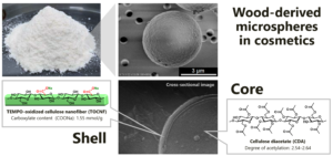
Cellulose nanofiber (CNF) derived from woody bioresources is a fascinating natural nanomaterial. In this work, spherical microparticles were fabricated by using TEMPO-oxidized CNF (TOCNF) and cellulose diacetate (CDA) via Pickering emulsion templating. The CDA-dissolved organic solvents were emulsified stably with TOCNF, followed by removing the solvents to form microspheres with core-shell structures, where the CDA cores were covered with the TOCNF shells. The prepared spherical microparticles possessed an average diameter and sphericity index of 6.4 μm and 0.96, respectively. The zeta-potential value was approximately -48 mV, indicating the stable colloidal system in water. The CDA/TOCNF microparticles were stained with toluidine blue dye for negatively-charged TOCNF. Besides, furry nanofiber-like morphology was observed on the particle surface by scanning electron microscopy. Wood-derived CDA/TOCNF microspheres are a promising alternative to fossil resource-derived, non-biodegradable microbeads in cosmetic applications.
Download PDF
Full Article
Preparation of Spherical Microparticles Composed of Cellulose Nanofiber and Cellulose Diacetate via Pickering Emulsion Templating
Yuna Tanaka,a Naoya Fukuda,a Naliharifetra Jessica Ranaivoarimanana,a,b Mayumi Hatakeyama,a and Takuya Kitaoka a,*
Cellulose nanofiber (CNF) derived from woody bioresources is a fascinating natural nanomaterial. In this work, spherical microparticles were fabricated by using TEMPO-oxidized CNF (TOCNF) and cellulose diacetate (CDA) via Pickering emulsion templating. The CDA-dissolved organic solvents were emulsified stably with TOCNF, followed by removing the solvents to form microspheres with core-shell structures, where the CDA cores were covered with the TOCNF shells. The prepared spherical microparticles possessed an average diameter and sphericity index of 6.4 μm and 0.96, respectively. The zeta-potential value was approximately -48 mV, indicating the stable colloidal system in water. The CDA/TOCNF microparticles were stained with toluidine blue dye for negatively-charged TOCNF. Besides, furry nanofiber-like morphology was observed on the particle surface by scanning electron microscopy. Wood-derived CDA/TOCNF microspheres are a promising alternative to fossil resource-derived, non-biodegradable microbeads in cosmetic applications.
DOI: 10.15376/biores.18.1.1482-1492
Keywords: TEMPO-oxidized cellulose nanofiber; Cellulose diacetate; Microsphere; Core-shell structure; Cosmetic applications
Contact information: a: Department of Agro-Environmental Sciences, Kyushu University, 744 Motooka, Nishi-ku, Fukuoka 819-0395, Japan; b: Yokohama Institute for Earth Sciences, Japan Agency for Marine-Earth Science and Technology, 3173-25 Showa-machi, Kanazawa-ku, Yokohama 236-0001, Japan.
* Corresponding author: tkitaoka@agr.kyushu-u.ac.jp
GRAPHICAL ABSTRACT
INTRODUCTION
A diverse array of plastics has long and widely enriched our daily life, but many of them cause environmental concerns due to their poor biodegradability and irreversible downsizing into micrometer-size pieces, called secondary microplastics (Browne et al. 2011; Cole et al. 2011). In addition, primary microplastics such as cosmetic microbeads are making matters worse because of impossible recovery once released into the ocean (Cheung and Fok 2017). The microplastics-derived oceanic pollution has brought serious damage to marine ecosystems, which threatens human survival (Auta et al. 2017). Therefore, an urgent solution is required.
Primary microplastics have sizes in micrometers. They are produced in large quantities from synthetic polymers, such as nylon and acrylics, in cosmetic applications for improving tactile sensation, and facial conditions (Cole et al. 2011). These small particles are difficult to recover once discharged, and they are not biodegradable in natural environments. Preferable alternatives to problematic plastic microbeads have been explored (Auta et al. 2017). One promising approach uses biodegradable synthetic polymers such as polylactide (PLA), polyhydroxybutyrate (PHB), and poly(ε-caprolactone) (PCL) (Slomkowski et al. 2005; Bokrova et al. 2019; Tasci et al. 2021). PLA is compostable but not biodegradable at all in water, while PHB and PCL are expected to degrade in the ocean (Briassoulis et al. 2020; Suzuki et al. 2021). The actual biodegradability of these polymers remains unknown due to the wide range of ocean conditions. The International Organization for Standardization (ISO) has proposed several laboratory-scale evaluation methods for biodegradation testing of plastics such as ISO 18830 (2016), ISO 22404 (2019), and ISO 19679 (2020), but the definition of marine biodegradability is now under consideration for international standardization. In any case, cosmetic microbeads need to be fabricated from abundant and biodegradable resources. In this view, PHB is produced in small quantity by bioengineered microorganisms, and PCL is a fossil fuel-based aliphatic polyester, resulting in strong demand for further refinements.
Cellulose, the main constituent of plant cell walls, is the most abundant biomass on the earth. It has a definite chemical structure, a linear chain of thousands of β(1→4) linked anhydro-D-glucopyranose unit, which self-assembles during biosynthesis to form a highly-crystalline nanofiber, called a cellulose nanofiber (CNF). Various CNF-based nanomaterials have been developed (Balea et al. 2021; Uetani et al. 2021; Hashemzehi et al. 2022; Szlek et al. 2022). This natural nano-polysaccharide is biodegraded promptly by microorganisms in the deep sea, such as marine bacterium Croceicoccus marinus (Shen et al. 2019). Cellulose acetate (CA) is a cellulose derivative, which has been produced from pure cellulose with acetic anhydride, and it is expected to be marine degradable as well as cellulose (Mazzotta et al. 2022). Acetyl groups are introduced at the C2, C3, and C6 positions of cellulose by esterification, and the degree of substitution (DS) affects biodegradability (Buchanan et al. 1993; Komarek et al. 1993; Sakai et al. 1996; Yadav and Hakkarainen 2022). Cellulose diacetate (CDA) microbeads have been launched on the market as a benign cosmetic additive, whose safety for living organisms has been confirmed by the American Society for Testing and Materials (ASTM D6691 (2018)).
Various types of CNF have attracted attention as nanomaterials obtained from woody bioresources, and the thixotropic property is expected to control the viscosity of cosmetic liquids (Goi et al. 2019). One of the unique properties of CNF involves amphiphilicity, showing spatially separated hydrophilic and hydrophobic faces (Glasser et al. 2012; Kalashnikova et al. 2012), which allows the formation of stable emulsions via CNF-mediated emulsification (Fujisawa et al. 2017; Kanomata et al. 2020). Surface-carboxylated CNF prepared by 2,2,6,6-tetramethylpiperidine 1-oxyl (TEMPO)-mediated oxidation possesses a large amount of carboxyl groups only at the C6 position of cellulose on the crystalline CNF surface, and allows the formation of a Pickering emulsion, which can efficiently homogenize oil and water phases due to electrostatic repulsion between stable Pickering emulsion particles (Kalashnikova et al. 2012; Goi et al. 2019). TEMPO-oxidized CNF (TOCNF) stabilized divinylbenzene droplets have been formed in an oil-in-water (o/w) Pickering emulsion, followed by in situ polymerization to form polydivinylbenzene microparticles covered with TOCNF (Fujisawa et al. 2019). The size and morphology of the obtained microparticles were suitable for cosmetic applications; however, the core polymers were non-biodegradable. Thus, challenges remain for marine biodegradability of cosmetic microparticles. A one-pot and one-step enzymatic synthesis of spherical microparticles composed of dehydrogenative polymers of coniferyl alcohol as a typical lignin precursor and TOCNF has been reported (Fukuda et al. 2021).
In this work, cellulose-derived microspheres were developed with a combination of TOCNF and CDA by oil-in-water (o/w) Pickering emulsion templating. Biodegradable CDA in the designated solvents was stably emulsified using an aqueous solution containing TOCNF via ultrasonic homogenization, followed by removing the solvents by simple washing treatment to form CDA cores. TOCNF acted as a solid surfactant of the Pickering emulsion, finally covering the CDA cores as a shell component. The cellulose-based core-shell microparticles fabricated from CDA and TOCNF are much different in the surface physicochemistry as compared to simple CDA microbeads, and they are expected to have potential for marine biodegradability.
EXPERIMENTAL
Materials
TOCNF used in this study was kindly provided by DKS Co. Ltd., Kyoto, Japan (RHEOCRYSTA, I-2SX, 2.3% (w/w), COONa = 1.55 mmol g-1). BELLOCEATM, commercial spherical microbeads prepared from CDA, was also kindly provided by Daicel Corporation, Osaka, Japan (average particle size: 7 µm). CDA (DS = 2.54 to 2.64) and cellulose triacetate (CTA, DS = 2.94 to 2.97) were purchased from FUJIFILM Wako Pure Chemical Industries, Ltd., Osaka, Japan. Pure-grade aniline, ethyl acetate, toluene and other solvents were purchased from Sigma-Aldrich Japan, Ltd., Tokyo, Japan, FUJIFILM Wako Pure Chemical Industries, Ltd., Osaka, Japan, and Tokyo Chemical Industry Co., Ltd., Tokyo, Japan. All other chemicals were of reagent grade and used as received without further purification. Water used in this study was purified using a Barnstead Smart2Pure system (Thermo Scientific Co. Ltd., Tokyo, Japan).
Methods
Characterization of TOCNF
Nanomorphology of TOCNF used in this study was observed by transmission electron microscopy (TEM; JEM-2100HCKM, JEOL Ltd., Tokyo, Japan) at an accelerating voltage of 200 kV. The diluted aqueous suspension (0.01 wt% TOCNF, 3 μL) was dropped onto a copper grid (elastic carbon coated, ELS-C10 STEM Cu100P grid specification, Ohken Shoji Co., Ltd., Tokyo, Japan), and then dyed with 1% sodium phosphotungstate for 3 min before the TEM observation. Atomic force microscopy (AFM; Dimension Icon, Bruker Japan Co. Ltd., Tokyo, Japan) equipped with a SCANASYST-AIR probe (k = 0.4 N m-1, F0 = 70 kHz) was applied for the shape measurement of TOCNF; 7 × 10⁻⁴ wt% of the TOCNF suspension was dropped on mica, and then air-dried. The measurement was performed with a scanning range of 2.0 × 2.0 µm2. Crystalline structure of TOCNF was recorded by X-ray diffraction (XRD) analysis using a Rigaku SmartLab diffractometer (Rigaku Co., Tokyo, Japan) with Ni-filtered Cu Kα radiation (λ = 0.15418 nm) at 40 kV and 20 mA. The scanning rate was 0.5° min-1 with 0.05° intervals. Carboxylate content of the TOCNF was determined by electrical conductivity titration method (Saito et al. 2006). TEM observations were performed at the Ultramicroscopy Research Center, Kyushu University. AFM and XRD analyses were conducted at the Center of Advanced Instrumental Analysis, Kyushu University.
Preparation of CDA/TOCNF Microparticles
CDA/TOCNF microparticles were prepared by the following method. CDA powder (2.4 g) was added to aniline (8 mL) or other solvents heated at 80 °C to achieve sufficient dissolution. After cooling down, TOCNF water suspension (0.8 wt%, 72 mL) was mixed with CDA/aniline, followed by vigorous sonication for 10 min using an ultrasonic homogenizer at 300 W and 20 kHz (US-300E, NIHONSEIKI KAISHA Ltd., Tokyo, Japan) to obtain stable o/w Pickering emulsion. The oil phase of the Pickering emulsion was gently removed by solvent diffusion using ethyl acetate/toluene mixture (1/1 by vol., 80 mL) for 15 min at room temperature.
This procedure was repeated four times using fresh washing solvents. After centrifugation at 12,000 G for 10 min, the supernatant was removed and further washed with water. The CDA/TOCNF microparticles were obtained by freeze-drying and passing through a sieve (φ = 212 µm). CTA was also used as a control to fabricate microparticles by Pickering emulsion templating. Schematic illustration for the preparation procedure is shown in Fig. 1.

Fig. 1. Preparation procedure of CDA/TOCNF microparticles via Pickering emulsion templating
Characterization of microparticles
As-prepared microparticles suspended in water (3 µL) were dropped on a carbon tape and air-dried in a desiccator at room temperature for 24 h. The dried samples were coated using an osmium coater (HPC-1SW, Vacuum Device, Ibaraki, Japan) for 1 s. Scanning electron microscopy (SEM, SU8000, Hitachi High-Tech Co, Tokyo, Japan) analysis was performed at 0.7 to 1.0 kV of an accelerating voltage.
The diameter and sphericity index of microparticle were calculated manually from the digital SEM images. In brief, each orthogonal minor axis was divided by the major axis for 176 microparticles. The microparticle samples embedded in a resin were sliced using an ultramicrotome (Ultracut-UCT, Leica, Wetzlar, Germany); the cross-section was observed by SEM observation, performed at the Center of Advanced Instrumental Analysis, Kyushu University.
Staining with toluidine blue dye for acidic polysaccharides was performed to characterize the TOCNF on the surface of microparticles. Thermogravimetric analysis (TG/DTA7300) was carried out at a heating rate of 5.0 °C min-1 from 50 to 500 °C under ambient conditions.
RESULTS AND DISCUSSION
Nanomorphology and Crystalline Structure of TOCNF
The morphology and crystalline structure of TOCNF used in this study are shown in Fig. 2. The average length and width were 406 ± 206 nm and 2.1 ± 0.7 nm, respectively. The width of the TOCNF, as reported ranging from 2 to 4 nm, was estimated by detecting the height of the TOCNF in the AFM measurement. Based on the low value of the observed height, some compaction might have occurred during the drying process. XRD analysis clearly showed a typical cellulose I crystalline structure, and the crystallinity was 61.8%, calculated by the Segal method (Segal et al. 1959). The carboxyl content of TOCNF used was 1.55 mmol g-1, determined by electrical conductivity titration. The obtained data for characterization of TOCNF were in good agreement with reported data (Saito et al. 2006). The TOCNF has been investigated as a solid surfactant to form Pickering emulsion, and thus the TOCNF was used for emulsification of CA solution in an oil-in-water systems.

Fig. 2. TEM image (a), AFM image (b), and XRD profile (c) of TOCNF used in this study
Preparation of Microparticles in Pickering Emulsion Systems
As illustrated in Fig. 1, water-immiscible solvents that can sufficiently dissolve CDA and CTA were required for making stable o/w Pickering emulsion. Ethyl acetate, methyl acetate, methyl pyrrolidone, and aniline were used in this study. Ethyl acetate and methyl acetate had a solubility of less than 1% for both CTA and CDA. Methyl pyrrolidone was able to dissolve CTA about 10% by weight; however, CTA precipitated when mixed with TOCNF suspension due to miscibility with water. Aniline was allowed to dissolve both CTA and CDA up to a concentration of 30%, which indicated that it is a promising solvent. However, 30% CTA/aniline solution had high viscosity, and it was difficult to sufficiently emulsify the solution by using aqueous TOCNF suspension. Lower concentration induced the unstable formation of microparticles during washing process with ethyl acetate. In contrast, 30% CDA/aniline solution could be homogeneously emulsified to form o/w Pickering emulsion with TOCNF. Commercial BELLOCEATM exhibited similar behavior to CTA, which was considered to possess higher DS value than CDA used in this study. Lower DS of CA is promising for higher marine degradability (Yadav and Hakkarainen 2022), and thus, 30% CDA/aniline solution was used to fabricate core components in this work.
The microparticles were fabricated during solvent removal from the Pickering emulsion, as illustrated in Fig. 1; therefore, the aniline-miscible solvents were required, e.g., ethyl acetate, toluene, and N,N-dimethylformamide (DMF). Spherical formation of CDA proceeded in the presence of ethyl acetate and toluene, although as-formed microparticles immediately dissolved by using DMF. Considering the ratio of ethyl acetate and toluene, a 1:1 mixture by volume was the best to obtain high yield (60.1%) without any agglomeration during precipitation. As-prepared samples were subjected to sufficient rinsing four times with a fresh ethyl acetate/toluene blend to remove any contaminants, and they were stable in the mixed solvent. After sieving with a 212-μm mesh, the yield dropped to 42.8%.
Morphological Characteristics of Pickering Emulsion and Microparticles
Ultrafine nanofibrous and crystalline TOCNF was used as a solid surfactant to form Pickering emulsion in this study. Figure 3a displays the powdery CDA/TOCNF composites fabricated by Pickering emulsion templating. Figure 3b shows well-dispersed droplets in water phase just after ultrasonic emulsification, and Fig. 3c exhibits spherical particles suspended in water after removal of solvents. SEM images visualize the spherical matters of as-prepared CDA/TOCNF microparticles, as shown in Fig. 3d and 3e. A size distribution histogram indicated that the average size of particles was 6.4 µm with a sphericity of 0.96 in Fig. 3f, although the relatively large particles were observed in Fig. 3d. Fine-sieving with a 20-μm mesh afforded 2.9% in the yield, which will require further improvement. Cosmetic applications of microparticles in general require the designated size of several micrometers in diameter; therefore, the CDA/TOCNF microspheres are expected to be used as cosmetic additives. The large variation in particle sizes was possibly due to the viscosity of liquids prior to emulsification.

Fig. 3. Visual (a) and optical microscopic images before (b) and after (c) solvent removal. SEM images of CDA/TOCNF microparticles (d, e). Size distribution histogram of the microparticles (f)
Core-shell Structure of CDA/TOCNF Microspheres
The CDA/TOCNF microspheres were stained with toluidine blue dye to characterize the surface of the particles. Toluidine blue well stained the CDA/TOCNF microparticles, although CDA without TOCNF remained almost unchanged after staining, as shown in Figs. 4a and 4b. Positively charged toluidine blue molecules presumably interacted with the carboxyl groups of TOCNF, resulting in clear light blue color. The characteristic metachromasy of toluidine blue from blue to purple-pink color via strong interaction with anionic polymer materials is well known, and was actually observed for the CDA/TOCNF suspension. However, the dried sample after washing exhibited light blue color as shown in Fig. 4a, due possibly to some aggregation of toluidine blue dye molecules. This result indicated that the surface of microparticles was covered with TOCNF. This conclusion was supported by the surface charge values. Table 1 lists the zeta-potential values of CDA/TOCNF microparticles and CDA particles without TOCNF, definitely indicating greater negative charge of CDA/TOCNF microparticles due to the dissociated carboxylate groups of TOCNF. From these results, higher dispersion stability in water is expected because of the strong electrostatic repulsion between microparticles.
Commercial BELLOCEATM has provided a moist feeling due to its hydrophobicity and poor water absorptivity. In contrast, as-prepared CDA/TOCNF microparticles demonstrated a smooth and dry feeling for human skin through moisture removal from the skin owing to hygroscopic TOCNF shells.

Fig. 4. Visual images of CDA/TOCNF microparticles (a) and CDA powders (b) stained with toluidine blue dye. (c) TGA profiles of TOCNF, CDA and CDA/TOCNF microparticle
Table 1. Zeta Potential Values of CDA/CNF Microparticles and CDA Powders

Cross-sectional SEM images of the CDA/TOCNF microparticle are shown in Fig. 5. The contrast difference between core and shell regions possibly indicated that a thin layer of TOCNF with furry appearance was located on the surface as a shell, and the core presumably consisted of different components from the shell. Furthermore, the core exhibited a dense structure without pores, which was distinctly different than the shaggy shell. To determine the amount of shell fraction ratio, TGA measurement was performed for CDA and TOCNF, which demonstrated the thermal decomposition temperatures of 340 °C and 214 °C, respectively, as shown in Fig. 4c. With reference to 280 °C between each pyrolysis point, the remaining weight of each component was 99.1% and 54.5%, respectively. CDA/TOCNF microparticles exhibited a residual weight of 96.4% at 280°C. Then, the TGA profiles of CDA and CDA/TOCNF seemed to be similar, possibly indicating that the major component of the CDA/TOCNF consisted of the CDA fraction. From the results, the shell weight fraction of the fine particles was estimated to be 6.0%, implying that the CDA/TOCNF microparticles possessed a thin surface layer. Therefore, the CDA/TOCNF microparticles presumably possessed the dense structure of CDA, as shown in Fig. 5.

Fig. 5. Cross-sectional SEM images of CDA/TOCNF microparticle (a) and the magnified surface (b)
The CDA/TOCNF microparticles with a core-shell structure are expected to degrade in the ocean, and further functionalization would be possible for loading and release of various functional cosmetic ingredients on the TOCNF layers. In the future work, hollow, spongy and biodegradable microspheres composed of CNF will be developed as a promising material that can carry various functional contents.
CONCLUSIONS
- Spherical fine particles of cellulose diacetate (CDA) and TEMPO-oxidized cellulose nanofiber (TOCNF) as a core and a shell, respectively, were successfully fabricated via Pickering emulsion templating.
- The microparticles demonstrated a smooth feeling for human skin and may fulfill requirements for cosmetic additives in terms of both particle size and sphericity.
ACKNOWLEDGMENTS
The authors are grateful for the supports of the JST-MIRAI program (Grant No. JPMJMI21EC) from Japan Science and Technology Agency (T.K.), and the Short-term Intensive Research Support Program from the Faculty of Agriculture, Kyushu University (M.H. and T.K.). We would like to thank Mr. Masaya Ohmura, DAICEL Corporation, for providing free samples of BELLOCEATM. The authors also appreciate technical assistance from the Ultramicroscopy Research Center, Kyushu University and the Center of Advanced Instrumental Analysis, Kyushu University.
REFERENCES CITED
ASTM D6691 (2018). “Standard test method for determining aerobic biodegradation of plastic materials in the marine environment by a defined microbial consortium or natural sea water inoculum,” ASTM International, West Conshohocken, PA.
Auta, H. S., Emenike, C. U., and Fauziah, S. H. (2017). “Distribution and importance of microplastics in the marine environment: A review of the sources, fate, effects, and potential solutions,” Environment International 102, 165-176. DOI: 10.1016/j.envint.2017.02.013
Balea, A., Blanco, A., Delgado-Aguilar, M., Concepcion Monte, M., Tarrés, Q., Fuente, E., Mutjé, P., and Negro, C. (2021). “Nanocellulose characterization challenges,” BioResources 16(2), 4382-4410. DOI: 10.15376/biores.16.2.Balea
Bokrova, J., Marova, I., Matouskova, P., and Pavelkova, R. (2019). “Fabrication of novel PHB-liposome nanoparticles and study of their toxicity in vitro,” Journal of Nanoparticle Research 21(3), 49. DOI: 10.1007/s11051-019-4484-7
Briassoulis, D., Pikasi, A., Papardaki, N. G., and Mistriotis, A. (2020). “Aerobic biodegradation of bio-based plastics in the seawater/sediment interface (sublittoral) marine environment of the coastal zone – Test method under controlled laboratory conditions,” Science of the Total Environment 722. DOI: 10.1016/j.scitotenv.2020.137748\
Browne, M. A., Crump, P., Niven, S. J., Teuten, E., Tonkin, A., Galloway, T., and Thompson, R. (2011). “Accumulation of microplastic on shorelines worldwide: Sources and sinks,” Environmental Science and Technology 45(21), 9175-9179. DOI: 10.1021/es201811s
Buchanan, C. M., Gardner, R. M., and Komarek, R. J. (1993). “Aerobic biodegradation of cellulose acetate,” Journal of Applied Polymer Science 47(10), 1709-1719. DOI: 10.1002/app.1993.070471001
Cheung, P. K., and Fok, L. (2017). “Characterisation of plastic microbeads in facial scrubs and their estimated emissions in Mainland China,” Water Research 122, 53-61. DOI: 10.1016/j.watres.2017.05.053
Cole, M., Lindeque, P., Halsband, C., and Galloway, T. S. (2011). “Microplastics as contaminants in the marine environment: A review,” Marine Pollution Bulletin. DOI: 10.1016/j.marpolbul.2011.09.025
Fujisawa, S., Togawa, E., and Kuroda, K. (2017). “Nanocellulose-stabilized Pickering emulsions and their applications,” Science and Technology of Advanced Materials. DOI: 10.1080/14686996.2017.1401423
Fujisawa, S., Togawa, E., Kuroda, K., Saito, T., and Isogai, A. (2019). “Fabrication of ultrathin nanocellulose shells on tough microparticles: Via an emulsion-templated colloidal assembly: Towards versatile carrier materials,” Nanoscale 11(32), 15004-15009. DOI: 10.1039/c9nr02612f
Fukuda, N., Hatakeyama, M., and Kitaoka, T. (2021). “Enzymatic preparation and characterization of spherical microparticles composed of artificial lignin and tempo-oxidized cellulose nanofiber,” Nanomaterials 11(4). DOI: 10.3390/nano11040917
Glasser, W. G., Atalla, R. H., Blackwell, J., Malcolm Brown, R., Burchard, W., French, A. D., Klemm, D. O., and Nishiyama, Y. (2012). “About the structure of cellulose: debating the Lindman hypothesis,” Cellulose 19, 589-598. DOI: 10.1007/s10570-012-9691-7
Goi, Y., Fujisawa, S., Saito, T., Yamane, K., Kuroda, K., and Isogai, A. (2019). “Dual functions of TEMPO-oxidized cellulose nanofibers in oil-in-water emulsions: A Pickering emulsifier and a unique dispersion stabilizer,” Langmuir 35(33), 10920-10926. DOI: 10.1021/acs.langmuir.9b01977
Hashemzehi, M., Mesic, B., Sjöstrand, B., and Naqvi, M. (2022). “A comprehensive review of nanocellulose modification and applications in papermaking and packaging: Challenges, technical solutions, and perspectives,” BioResources. DOI: 10.15376/biores.17.2.Hashemzehi
ISO 18830 (2016). “Plastics – Determination of aerobic biodegradation of non-floating plastic materials in a seawater/sandy sediment interface – Method by measuring the oxygen demand in closed respirometer,” International Organization for Standardization, Geneva, Switzerland.
ISO 22404 (2019). “Plastics – Determination of the aerobic biodegradation of non-floating materials exposed to marine sediment – Method by analysis of evolved carbon dioxide,” International Organization for Standardization, Geneva, Switzerland.
ISO 19679 (2020). “Plastics – Determination of aerobic biodegradation of non-floating plastic materials in a seawater/sediment interface – Method by analysis of evolved carbon dioxide,” International Organization for Standardization, Geneva, Switzerland.
Kalashnikova, I., Bizot, H., Cathala, B., and Capron, I. (2012). “Modulation of cellulose nanocrystals amphiphilic properties to stabilize oil/water interface,” Biomacromolecules 13(1), 267-275. DOI: 10.1021/bm201599j
Kanomata, K., Fukuda, N., Miyata, T., Lam, P. Y., Takano, T., Tobimatsu, Y., and Kitaoka, T. (2020). “Lignin-inspired surface modification of nanocellulose by enzyme-catalyzed radical coupling of coniferyl alcohol in Pickering emulsion,” ACS Sustainable Chemistry and Engineering 8(2), 1185-1194. DOI: 10.1021/acssuschemeng.9b06291
Komarek, R. J., Gardner, R. M., Buchanan, C. M., and Gedon, S. (1993). “Biodegradation of radiolabeled cellulose acetate and cellulose propionate,” Journal of Applied Polymer Science 50(10), 1739-1746. DOI: 10.1002/app.1993.070501009
Mazzotta, M. G., Reddy, C. M., and Ward, C. P. (2022). “Rapid degradation of cellulose diacetate by marine microbes,” Environmental Science and Technology Letters, 9(1), 37-41. DOI: 10.1021/acs.estlett.1c00843
Saito, T., Nishiyama, Y., Putaux, J. L., Vignon, M., and Isogai, A. (2006). “Homogeneous suspensions of individualized microfibrils from TEMPO-catalyzed oxidation of native cellulose,” Biomacromolecules 7(6), 1687-1691. DOI: 10.1021/bm060154s
Sakai, K., Yamauchi, T., Nakasu, F., and Ohe, T. (1996). “Biodegradation of cellulose acetate by Neisseria sicca,” Biosci. Biotech. Biochem. 60(10), 1617-1622. DOI: 10.1271/bbb.60.1617
Segal, L., Creely, J. J., Martin, A. E., and Conrad, C. M. (1959). “An empirical method for estimating the degree of crystallinity of native cellulose using the X-ray diffractometer,” Textile Research Journal 29(10), 786-794. DOI: 10.1177/004051755902901003
Shen, Y., Li, Z., Huo, Y. Y., Bao, L., Gao, B., Xiao, P., Hu, X., Xu, X. W., and Li, J. (2019). “Structural and functional insights into CmGH1, a novel GH39 family β-glucosidase from deep-sea bacterium,” Frontiers in Microbiology 10. DOI: 10.3389/fmicb.2019.02922
Slomkowski, S., Gadzinowski, M., Sosnowski, S., de Vita, C., Pucci, A., Ciardelli, F., Jakubowski, W., and Matyjaszewski, K. (2005). “Biodegradable nano- and microparticles with controlled surface properties,” Macromol. Symp., 226, 239-252. DOI: 10.1002/masy.200550822
Suzuki, M., Tachibana, Y., and Kasuya, K. I. (2021). “Biodegradability of poly(3-hydroxyalkanoate) and poly(ε-caprolactone) via biological carbon cycles in marine environments,” Polymer Journal 53, 47-66. DOI: 10.1038/s41428-020-00396-5
Szlek, D. B., Reynolds, A. M., and Hubbe, M. A. (2022). “Hydrophobic molecular treatments of cellulose-based or other polysaccharide barrier layers for sustainable food packaging: A review,” BioResources 17(2), 3551-3673. DOI: 10.15376/biores.17.2.Szlek
Tasci, M. E., Dede, B., Tabak, E., Gur, A., Sulutas, R. B., Cesur, S., Ilhan, E., Lin, C. C., Paik, P., Ficai, D., Ficai, A., and Gunduz, O. (2021). “Production, optimization and characterization of polylactic acid microparticles using electrospray with porous structure,” Applied Sciences (Switzerland) 11(11). DOI: 10.3390/app11115090
Uetani, K., Ranaivoarimanana, N. J., Hatakeyama, M., and Kitaoka, T. (2021). “Inherently distinctive potentialities and uses of nanocellulose based on its nanoarchitecture,” BioResources 16(2), 4438-4473. DOI: 10.15376/biores.16.2.4438-4473
Yadav, N., and Hakkarainen, M. (2022). “Degradation of cellulose acetate in simulated aqueous environments: One-year study,” Macromolecular Materials and Engineering 307(6). DOI: 10.1002/mame.202100951
Article submitted: November 25, 2022; Peer review completed: December 21, 2022; Revised version received and accepted: December 24, 2022; Published: January 9, 2023.
DOI: 10.15376/biores.18.1.1482-1492